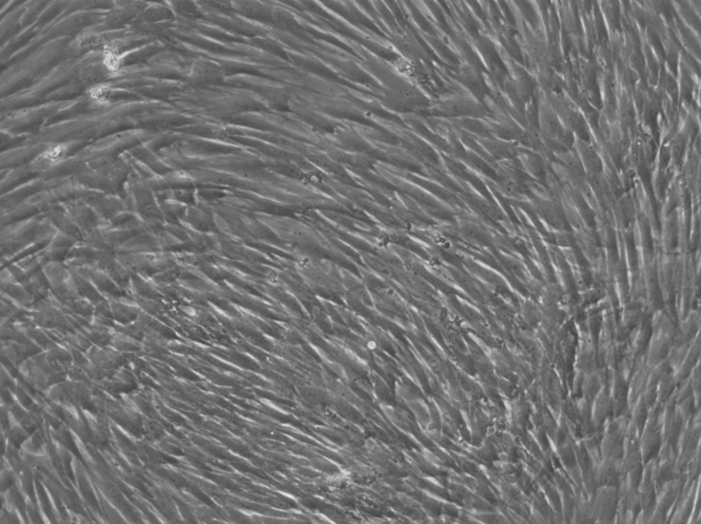
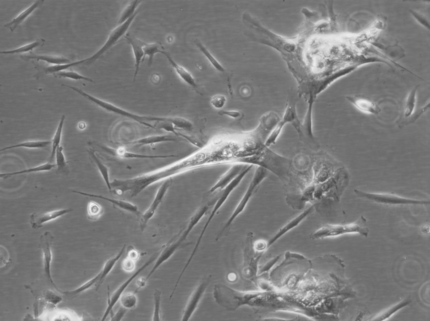
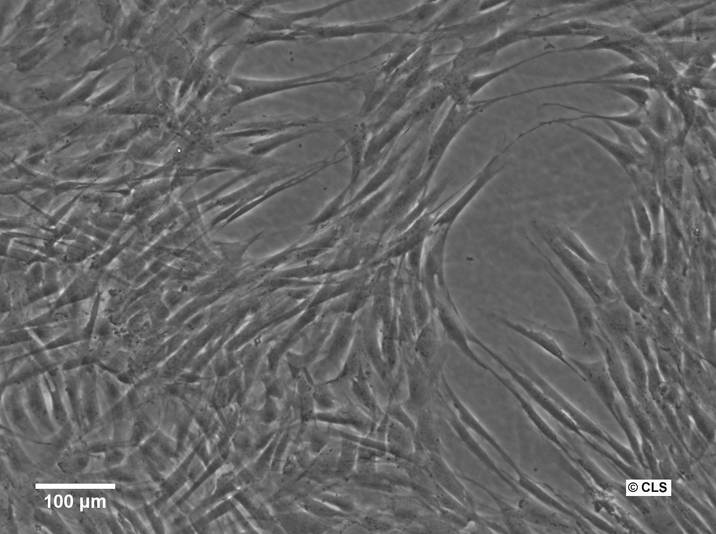
Human Primary Cells at Cytion: Specialized and Diverse for Advanced Research
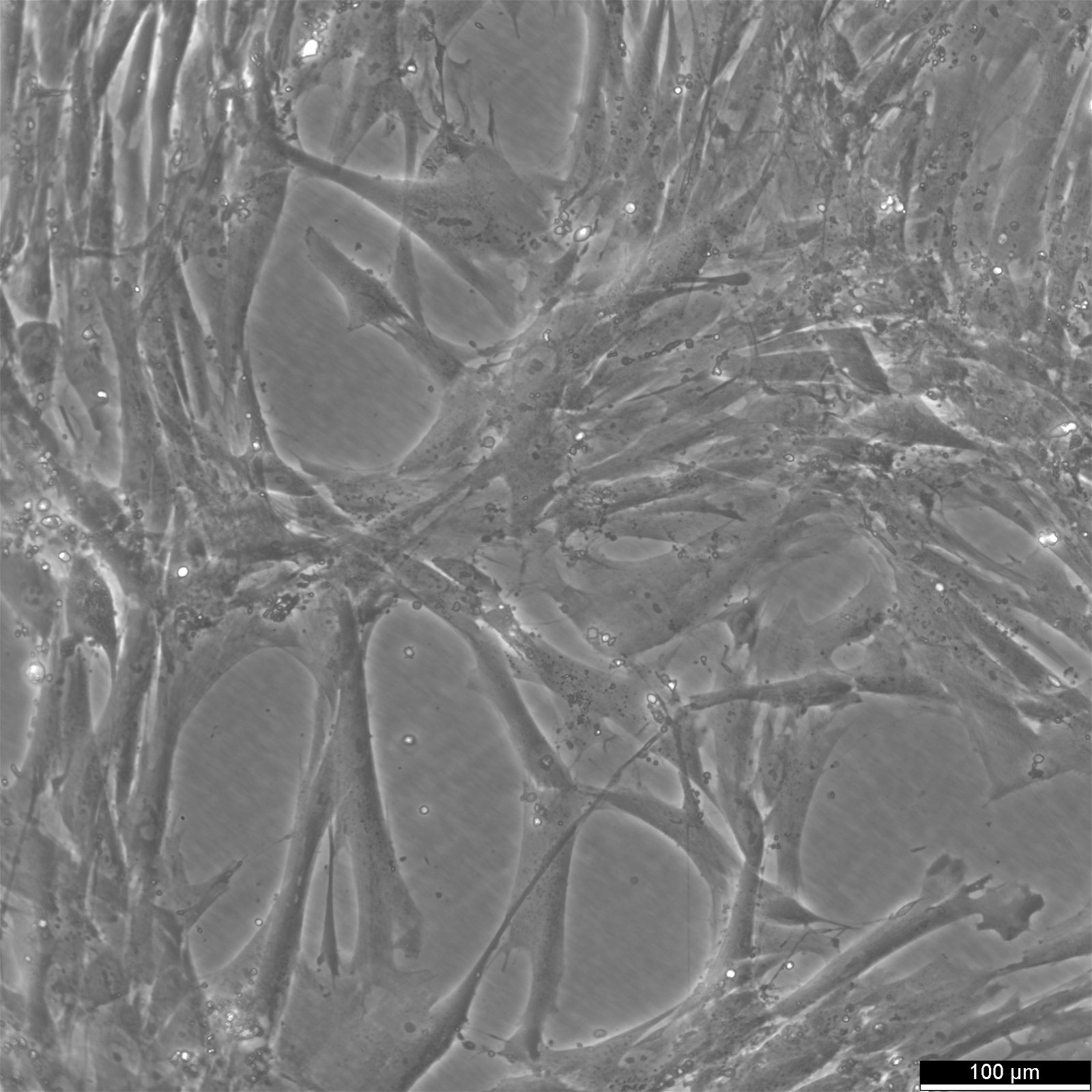
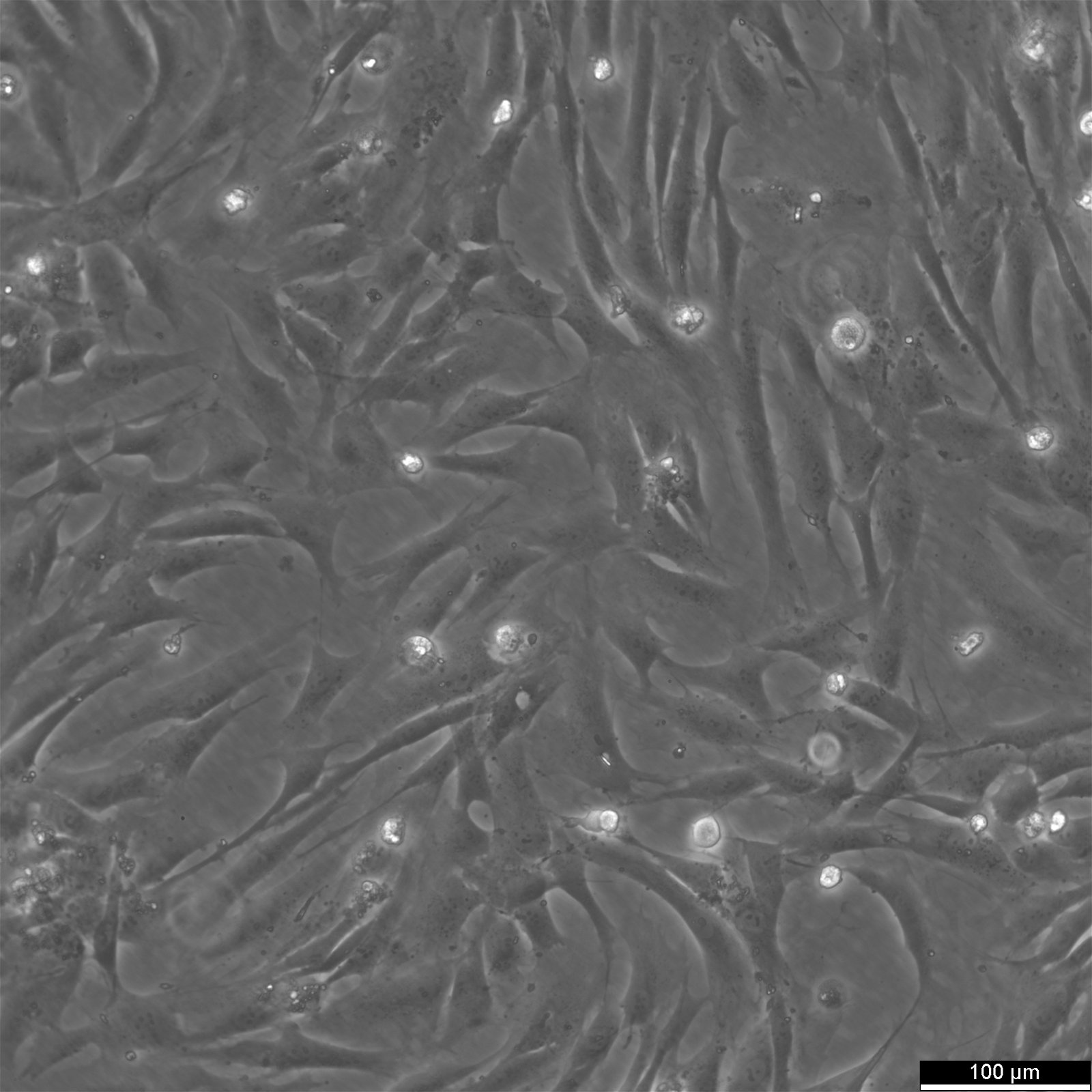
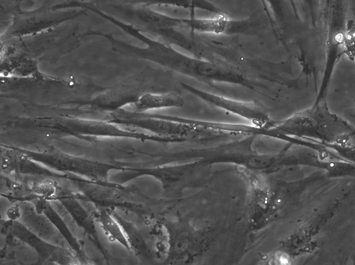
Explore Cytion's diverse range of human primary cells, meticulously chosen to support cutting-edge scientific inquiry. Our selection includes specialized cell types such as HUVECs (Human Umbilical Vein Endothelial Cells), Human Dental Follicle Stem Cells (hDFSC), and Human Gingival Fibroblasts (hGF).
HUVECs, sourced from single donors, are essential for vascular biology and angiogenesis studies. hDFSCs, vital for dental and craniofacial research, and hGFs, pivotal in oral biology and wound healing studies, reflect our commitment to providing high-quality, physiologically relevant cells. Each cell type is authenticated and rigorously tested, ensuring purity and viability for impactful research outcomes.
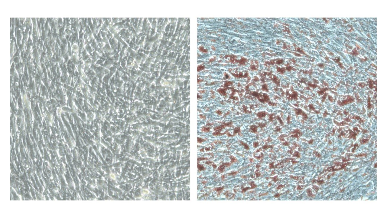
What are human primary cells?
Primary cells are the purest representation of their respective tissues. They are isolated from the tissue and processed so that they can become established in a culture setting with ideal conditions. They more closely mimic the in vivo state and display normal physiology because they are derived from tissue rather than modified. Because of this, they can serve as useful models for research into cellular pharmacology, toxicology, and physiology (including studies of metabolism, aging, and signal transduction). Keep in mind that primary cells are more challenging to culture and maintain than a continuous cell line because they have a shorter lifespan and will stop dividing (or senesce) after a certain number of cell divisions. Studies of cell signaling pathways are complicated by the inherent variability of primary cells acquired from donors and through subculture practices. Before beginning signaling studies, researchers often conduct a screening to determine whether or not the cells respond to commonly used stimuli. To avoid wasting time and money, primary cells can be stimulated to activate major signaling pathways before being screened.
Why use human primary cells?
Immortalized cell lines are commonly used as a cell assay. Although scientists have acknowledged that biological changes due to cell lines may be harmful in studying their physiological significance. The use of human primary cells improves the physiologic value of data obtained through cell cultures, and they are increasingly regarded as being important for studying biological processes, disease progress, and drug development.
Human primary cells are widely used in in vitro studies of intercellular and intracellular communication, developmental biology, and the mechanisms underlying cancer, Parkinson's disease, and diabetes, among many other preclinical and investigative biological research areas. Researchers have long used immortalized cell lines to study tissue function; however, cell lines with obvious mutations and chromosomal abnormalities may not be good surrogates for normal cells and the development of disease in its early stages. A more accurate model of a specific tissue cell type can now be achieved by using human primary cells isolated from that tissue and maintained in primary cell culture media and supplements.
What is primary cell culture?
Instead of using immortalized cell lines, primary cell culture involves growing cells directly from a multicellular organism outside of the body. Legal recognition exists in some countries, such as the UK, for the fact that primary cell cultures are more representative of in vivo tissues than cell lines. Nonetheless, primary cells need the right substrate and nutrients to grow, and after a certain number of divisions, they develop a senescent phenotype that causes them to permanently stop dividing. These two factors motivate the creation of cell lines. Both naturally immortalized primary cells (e.g., HeLa cells) and artificially immortalized primary cells (e.g., HEK cells) can be cultured indefinitely in cell culture.
Human primary cells by tissue types
Epithelial cells, fibroblasts, keratinocytes, melanocytes, endothelial cells, muscle cells, immune cells, and stem cells like mesenchymal stem cells are among the most commonly used human primary cells in scientific study. To begin with, the cultures are heterogeneous (representing a mix of cell types present in the tissue), and they can only be kept alive in vitro for a specific amount of time. Transformation is an in vitro process that allows human primary cells to be manipulated for unlimited subcultures. Transforming can happen naturally, or it can be induced by chemicals or viruses. After undergoing genetic transformation, a primary culture can divide indefinitely into an immortalized secondary cell line if given enough nutrients and space.
Endothelial Cells
Cancer treatment, wound healing, cell signaling research, high-throughput and high-content screening, and toxicology screening are just some of the areas that can benefit from the use of primary endothelial cells as a research tool.
Keratinocytes
Keratinocytes, derived from the epidermis of either adult human skin or neonatal foreskin, play a crucial role in the study of skin diseases like psoriasis and cancer.
Epithelial Cells
From cancer studies to toxicological investigations, primary epithelial cells have proven to be invaluable resources for modeling the body's natural defenses.
Fibroblasts
Inducing pluripotent stem (iPS) cells and studying wound healing are just a few of the many uses for primary fibroblasts.
Immune cells
Peripheral blood mononuclear cells, PBMC for short, are mononuclear cells of the blood with a round cell nucleus. They mainly include lymphocytes and monocytes, which take on important functions in the course of an immune response. Peripheral blood mononuclear cells are often used to diagnose infections or to detect possible vaccination protection. Insight into the cellular immune response mediated by T cells is often crucial.
Melanocytes
Melanocytes, the specialized skin cells that produce the pigment melanin, are helpful as models for research into topics such as wound healing, toxicity, melanoma, the dermal response to ultraviolet (UV) radiation, skin diseases, and cosmetics.
Stem cells
Stem cells have the potential to differentiate into a wide variety of cell types. Due to their ability to differentiate, they provide fresh opportunities for modeling human tissue and health conditions.
Mesenchymal stem cells
Mesenchymal stem cells, also known as MSCs, may be obtained from different human sources like bone marrow, fat (adipose tissue), umbilical cord tissue (Wharton's Jelly), and amniotic fluid (the fluid surrounding a fetus) and can be expanded in vitro. These adult stromal stem cells have the ability to develop into a wide variety of cell types. Some of these cell types include bone cells, cartilage cells, muscle cells, neural cells, skin cells, and corneal cells.
Smooth muscle cells
Within hollow organs, primary smooth muscle cells (SMCs) line the interior and mediate contractility. In addition to cancer and other diseases, SMCs can be used to model hypertension fibrosis.
Primary cells and cell lines
Either by spontaneous mutation, as in transformed cancer cell lines, or through intentional alteration, as in the artificial production of cancer genes, continuous cell lines have gained the potential to reproduce endlessly (immortalized). As a rule, continuous cell lines are more reliable and convenient to deal with than primary cells. They can expand indefinitely and provide rapid access to essential data. The use of continuous cell lines has certain limitations, including the fact that they are genetically modified/transformed, which might change physiological features and not match the in vivo condition, and that this can further change over time with significant passaging.
Advances in primary cell culture
Primary cells have a notorious reputation for being difficult to work with. The process, however, is becoming easier than ever before thanks to developments in primary cell culture, the availability of commercial primary cells with fully optimized protocols, and new analysis techniques that require less input.
The shift from two-dimensional to three-dimensional cell culture is regarded as a major milestone in the field. Tissue-specific architecture, cell-cell interactions, and mechanical/biochemical signaling may be attenuated in a 2D culture. Thus, there is a ceiling to the biological value of these cultures.
On the other hand, 3D cell culture enables cells to expand and interact with a 3D extracellular framework. This allows cells to interact with each other and the extracellular matrix, making 3D cultures more physiologically relevant. This method's accuracy in predicting in vivo responses has made it revolutionary in fields like drug discovery and development. Because of this, state-of-the-art technologies, like organoids derived from patients and organs-on-a-chip, provide highly contextual models for drug screening and development.
Primary cell generation is a bottleneck in primary culture. A larger volume of tissue is usually required to overcome this, which can be challenging to attain. However, improved analytical sensitivity is providing a way forward. For example, the need to culture large quantities of primary cells is reduced by using single-cell technology, which includes sequencing, western blotting, and mass cytometry.
Promising prospects for primary cell culture
The overall difficulties of primary cell culture are being mitigated by technological advances. In turn, this method is rapidly replacing others as the gold standard in cellular and molecular biology study and practice. Vaccine manufacturing, organ replacement, stem cell therapies, cancer research, and so much more stand to benefit greatly from the continued advancements in primary cell culture.
Primary cell culture tips and tricks
The needs of cell expansion
The two most common methods for cultivating primary cells are in suspension or on a surface (2D). Some cells are able to float freely in the bloodstream without ever becoming adherent to a surface (for example those derived from peripheral blood). Different cell lines have been engineered to thrive in suspension cultures, where they can reach densities unattainable under 2D growth conditions. Primary cells that need anchorage to grow in vitro are called adherent cells and include those found in solid tissues. To improve adhesion properties and supply other signals required for growth and differentiation, these cells are typically cultured in a flat uncoated plastic vessel, but occasionally a micro-carrier. This latter option may be coated with extracellular matrix proteins (such as collagen and laminin). The media used in cell culture consists of a basic medium that has been supplemented with the proper growth factors and cytokines. A cell incubator is a special type of laboratory incubator used to cultivate and maintain cells at a specific temperature and gas mixture (typically 37 °C, 5% CO2 for mammalian cells). Dependent on the type of cell being cultured, the optimal conditions can be very different. Depending on the types of cells being grown, the optimal growth medium will have a unique combination of factors, including but not limited to pH, glucose concentration, growth factors, and the presence of other nutrients.
Antibiotics in the growth medium are crucial during primary culture establishment to prevent contamination from the host tissue. Some antibiotic regimens feature a combination of gentamicin, penicillin, streptomycin, and amphotericin B. The use of antibiotics for an extended period of time is not advised, however, because some reagents (such as amphotericin B) may be toxic to cells in the long run.
Most primary cells go through senescence and stop dividing after a certain number of population doublings, making it crucial to keep them alive after isolation. Long-term cell viability requires expert cell-culturing techniques and ideal culture conditions (including the right medium, the right temperature, the right gas mixture, the right pH, the right concentration of growth factors, the presence of nutrients, and the presence of glucose). Since many of the growth factors used to supplement media are obtained from animal blood (the blood-derived ingredients possess the potential for contamination), it is recommended that their use be minimized or avoided altogether. It's also important to use an aseptic technique.
Subculture and maintenance
When cells in isolation adhere to the surface of the culture dish, this marks the beginning of the maintenance phase. Attachment typically occurs 24 hours after cultural initiation. Cells should be subcultured when they have reached a certain confluence percentage and are actively replicating. Since post-confluent cells may undergo differentiation and exhibit slower proliferation after passage, it is best to subculture primary cell cultures before they reach 100% confluence.
Sub-culturing in fresh media maintains the exponential growth of anchorage-dependent cells. Subculturing monolayers disrupt inter- and intracellular cell-surface interactions. Low concentrations of proteolytic enzymes, such as trypsin/EDTA, are employed to extract adherent primary cells from monolayers or tissues. After dissociated and diluted into a single-cell solution, the cells are counted and transferred to fresh culture containers to re-attach and multiply.
Cryopreservation and recovery
Cryopreservation preserves live cells by freezing them at low temperatures. Cryopreserving and thawing human primary cells prevents cell death and damage during storage and usage. Human primary cells are cryoprotected using DMSO or glycerol (at the correct temperature and with a controlled rate of freezing). The freezing process must be progressive, at -1 °C each minute, to prevent ice crystal formation. Long-term storage requires liquid nitrogen (-196 °C) or temperatures below -130 °C.
Submerging frozen cells in a 37 °C water bath for about 1 to 2 minutes is all it takes to thaw cryopreserved cells. Human primary cells should not be centrifuged after thawing out of the freezer (as they are extremely sensitive to damage during recovery from cryopreservation). It's suitable for plating cells immediately after thawing, and it promotes attachment in cultures during the first 24 hours after plating. 1 After the cryopreserved primary cells have attached, the spent media must be removed (as DMSO is harmful to primary cells and may cause a drop in post-thaw viability).

