3T3-L1 Cells


Key points about the 3T3-L1 cell line
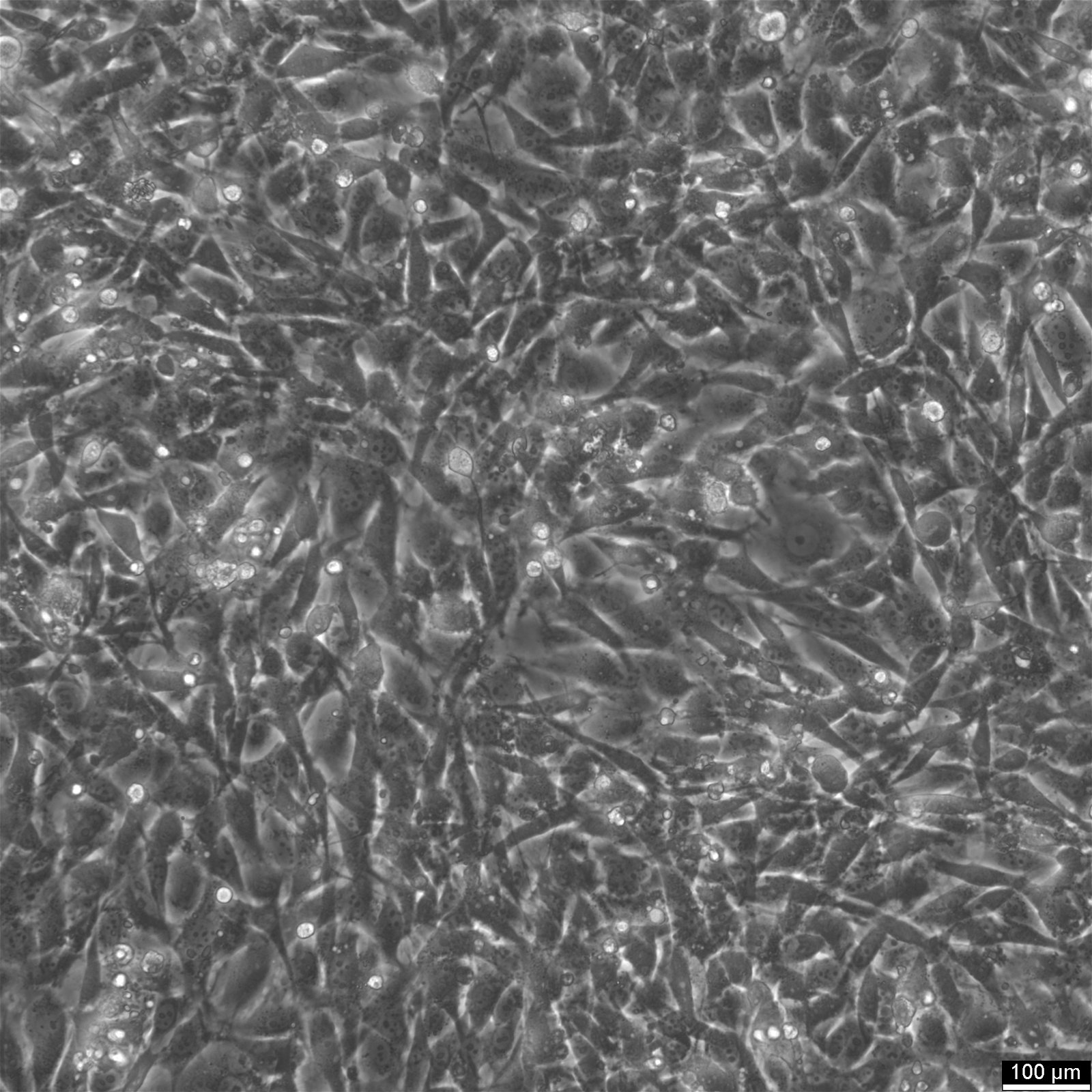
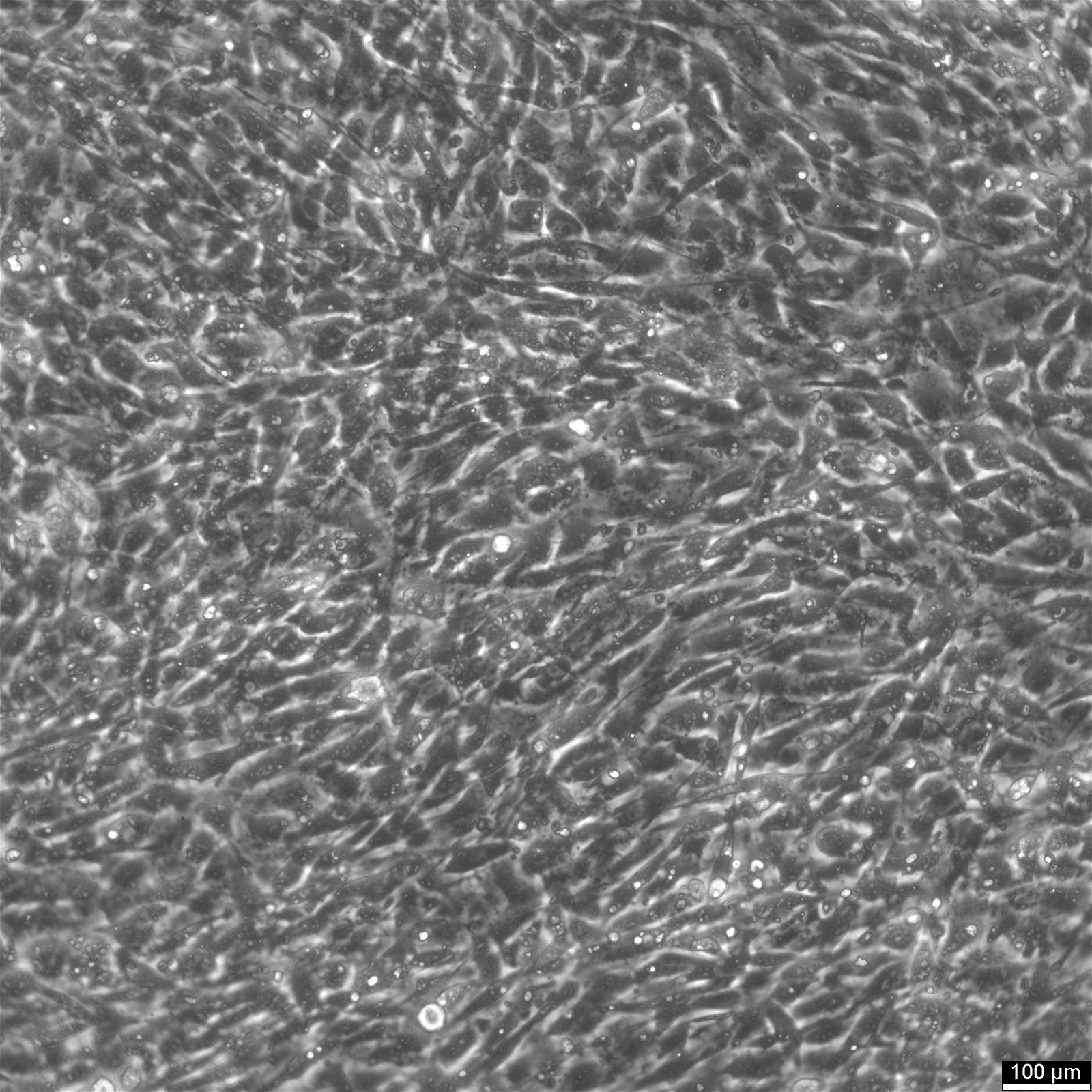
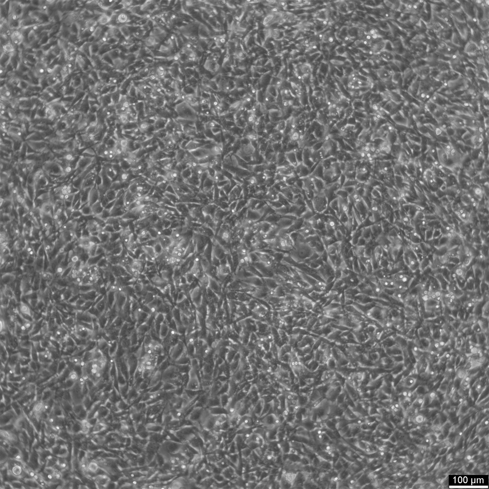
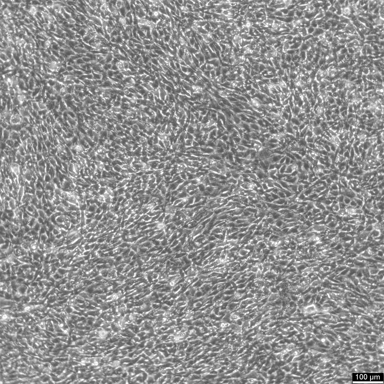
| Description | 3T3-L1 cells are a clonal line of preadipocytes derived from mouse embryonic fibroblasts. These cells have become a widely used in vitro model for studying the process of adipogenesis, including adipogenesis and lipogenesis, which is the differentiation of preadipocytes into adipocytes (fat cells). The name "3T3" refers to the transfer (T) protocol that involved transferring the cells every 3 days, and "L1" signifies the particular clone that was isolated. Initially, 3T3-L1 cells exhibit a fibroblast-like morphology, but upon induction of 3T3-L1 cell differentiation, 3T3-L1 cells change from a preadipocyte to a mature adipocyte state and accumulate lipid droplets, a hallmark of obesity and metabolic syndrome. The differentiation process from 3T3-L1 preadipocytes to 3T3-L1 adipocytes is triggered by a specific cocktail of inducers, typically including dexamethasone, 3-isobutyl-1-methylxanthine (IBMX), and insulin. As 3T3-L1 adipocytes adopt the characteristics of mature adipocytes, they begin to express genes that are crucial for adipocyte function, such as those coding for enzymes involved in fatty acid metabolism and hormones like leptin and adiponectin, which play vital roles in regulating appetite, energy balance, and insulin sensitivity. Studying 3T3-L1 cell transformations enhances our understanding of adipogenesis and obesity and fat-related diseases, such as type 2 diabetes, by revealing how lipid accumulation in adipocytes leads to cellular dysfunction and broader metabolic issues. Moreover, the 3T3-L1 cell line is instrumental in investigating the impact of various substances on adipocyte behavior, such as the effect of pharmacological agents on lipolysis or the anti-inflammatory properties of certain diets that may prevent insulin resistance. 3T3-L1 cells have been extensively used to study the molecular and cellular mechanisms underlying adipocyte differentiation, insulin sensitivity, lipid metabolism, and the effects of various nutritional and pharmacological agents on these processes. Given their ability to differentiate into adipocytes and their ease of culture in vitro, 3T3-L1 cells provide a valuable model system for obesity and diabetes research, as well as for the discovery of new therapeutic targets related to metabolic dise |
|---|---|
| Organism | Mouse |
| Tissue | Embryonic |
| Applications | 3T3-L1 cells have been used as a model system for understanding the molecular mechanisms that regulate adipogenesis and lipid metabolism, and have been utilized in research related to obesity, diabetes, and metabolic diseases. They are also a viable transfection host. |
| Synonyms | 3T3 L1, 3T3L1, 3T3-L1 ad, NIH-3T3-L1, NIH3T3-L1 |
Aspects
| Age | Embryo |
|---|---|
| Gender | Male |
| Morphology | Fibroblast-like |
| Growth properties | Adherent |
Specifications
| Citation | 3T3-L1 (Cytion catalog number 400103) |
|---|---|
| Biosafety level | 1 |
Genetic makeup of cell line 3T3-L1
| Tumorigenic | No |
|---|---|
| Virus susceptibility | murine leukemia virus, murine sarcoma virus, vesicular stomatitis, vaccinia, herpes simplex, N-tropic oncornaviruses C |
| Products | Insulin, collagen, triglycerides |
| Ploidy status | Aneuploid |
| Karyotype | 2n=40 |
Handling of 3T3-L1
| Culture Medium | DMEM, w: 4.5 g/L Glucose, w: 4 mM L-Glutamine, w: 1.5 g/L NaHCO3, w: 1.0 mM Sodium pyruvate (Cytion article number 820300a) |
|---|---|
| Medium supplements | Supplement the medium with 10% FBS |
| Passaging solution | Accutase |
| Subculturing | Remove the old medium from the adherent cells and wash them with PBS that lacks calcium and magnesium. For T25 flasks, use 3-5 ml of PBS, and for T75 flasks, use 5-10 ml. Then, cover the cells completely with Accutase, using 1-2 ml for T25 flasks and 2.5 ml for T75 flasks. Let the cells incubate at room temperature for 8-10 minutes to detach them. After incubation, gently mix the cells with 10 ml of medium to resuspend them, then centrifuge at 300xg for 3 minutes. Discard the supernatant, resuspend the cells in fresh medium, and transfer them into new flasks that already contain fresh medium. |
| Freeze medium | CM-1 (Cytion catalog number 800100) or CM-ACF (Cytion catalog number 806100) |
| Handling of cryopreserved cultures |
|
3T3 L1 cell purity and identity checks
| Sterility | Mycoplasma contamination is excluded using both PCR-based assays and luminescence-based mycoplasma detection methods. To ensure there is no bacterial, fungal, or yeast contamination, cell cultures are subjected to daily visual inspections. |
|---|
Required products
- A Gentle Alternative to Trypsin
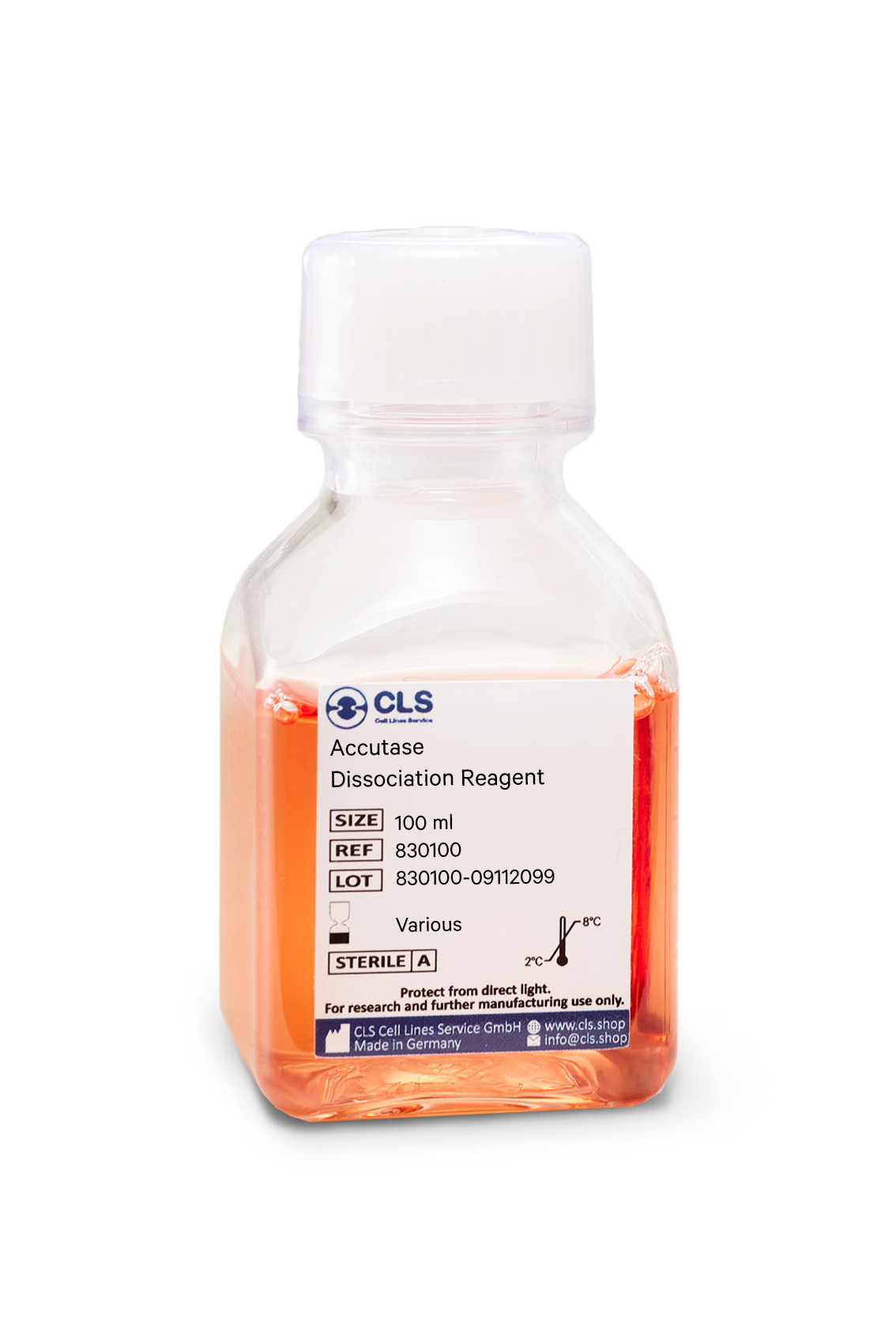
Accutase is a cell detachment solution that is revolutionizing the cell culture industry. It is a mix of proteolytic and collagenolytic enzymes that mimics the action of trypsin and collagenase. Unlike trypsin, Accutase does not contain any mammalian or bacterial components and is much gentler on cells, making it an ideal solution for the routine detachment of cells from standard tissue culture plasticware and adhesion coated plasticware. In this blog post, we will explore the benefits and uses of Accutase and how it is changing the game in cell culture.
Advantages of Accutase
Accutase has several advantages over traditional trypsin solutions. Firstly, it can be used whenever gentle and efficient detachment of any adherent cell line is needed, making it a direct replacement for trypsin. Secondly, Accutase works extremely well on embryonic and neuronal stem cells, and it has been shown to maintain the viability of these cells after passaging. Thirdly, Accutase preserves most epitopes for subsequent flow cytometry analysis, making it ideal for cell surface marker analysis.
Additionally, Accutase does not need to be neutralized when passaging adherent cells. The addition of more media after the cells are split dilutes Accutase so it is no longer able to detach cells. This eliminates the need for an inactivation step and saves time for cell culture technicians. Finally, Accutase does not need to be aliquoted, and a bottle is stable in the refrigerator for 2 months.
Applications of Accutase
Accutase is a direct replacement for trypsin solution and can be used for the passaging of cell lines. Additionally, Accutase performs well when detaching cells for the analysis of many cell surface markers using flow cytometry and for cell sorting. Other downstream applications of Accutase treatment include analysis of cell surface markers, virus growth assay, cell proliferation, tumor cell migration assays, routine cell passage, production scale-up (bioreactor), and flow cytometry.
Composition of Accutase
Accutase contains no mammalian or bacterial components and is a natural enzyme mixture with proteolytic and collagenolytic enzyme activity. It is formulated at a much lower concentration than trypsin and collagenase, making it less toxic and gentler, but just as effective.
Efficiency of Accutase
Accutase has been shown to be efficient in detaching primary and stem cells and maintaining high cell viability compared to animal origin enzymes such as trypsin. 100% of cells are recovered after 10 minutes, and there is no harm in leaving cells in Accutase for up to 45 minutes, thanks to autodigestion of Accutase.
In summary
In conclusion, Accutase is a powerful solution that is changing the game in cell culture. With its gentle nature, efficiency, and versatility, Accutase is the ideal alternative to trypsin. If you are looking for a reliable and efficient solution for cell detachment, Accutase is the solution for you.
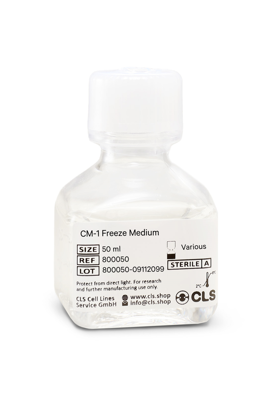
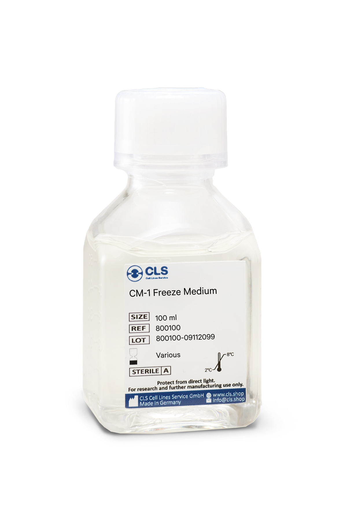
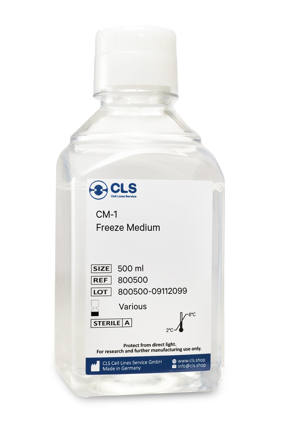
In biological research, the cryopreservation of mammalian cells is an invaluable tool. Successful preservation of cells is a top priority given that losing a cell line to contamination or improper storage conditions leads to lost time and money, ultimately delaying research results. Once the cells have been transferred from a cell growth medium to a freezing medium, the cells are typically frozen at a regulated rate and stored in liquid nitrogen vapor or at below -130°C in a mechanical deep freezer. The freeze medium CM-1 enables cryopreservation of cells at below -130°C (or in liquid nitrogen), essentially eliminating the need for an additional, costly ultralow freezer and eliminating time-consuming and demanding controlled rate freezing processes. Simply collect the cells, aspirate the growth medium, resuspend in CM-1, transfer to a cryovial, and store the vial at below -130 °C.
Long shelf-life
CM-1 is a serum-containing, ready-to-use cryopreservation medium that can be stored in the refrigerator for up to one year.
Trusted by hundreds of researchers
Our advanced cell freezing medium CM-1 is a market-leading product in Germany and Europe and is distinguished by numerous publications involving hundreds of different cell lines worldwide. We tested it with more than 1000 cell lines from our proprietary cell bank.
Optimized ingredients
CM-1 does contain serum products. Serum-containing cryopreservation mediums optimally protect the cells whilst being frozen and have the advantage of high recovery rates. As CM-1 has been tested with a multitude of cell lines, you can rest assured that your cells always recover well.
Contains FBS, DMSO, glucose, salts
Buffering capacity pH = 7.2 to 7.6
Applications & Validation
The cells preserved in our CM-1 freeze medium can be used for cell counting, viability and cryopreservation, cell culture, mammalian cell culture, gene expression analysis and genotyping, in vitro transcription, and polymerase chain reactions. Each batch's efficacy is evaluated using CHO-K1 cells. Each batch is tested for pH, osmolality, sterility, and endotoxins to ensure high quality.
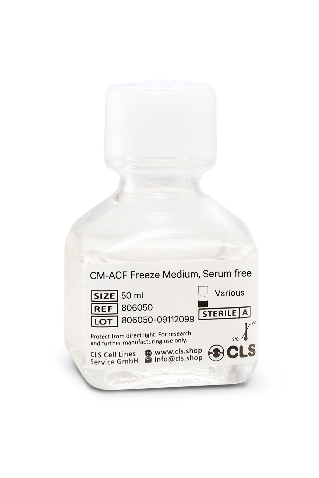
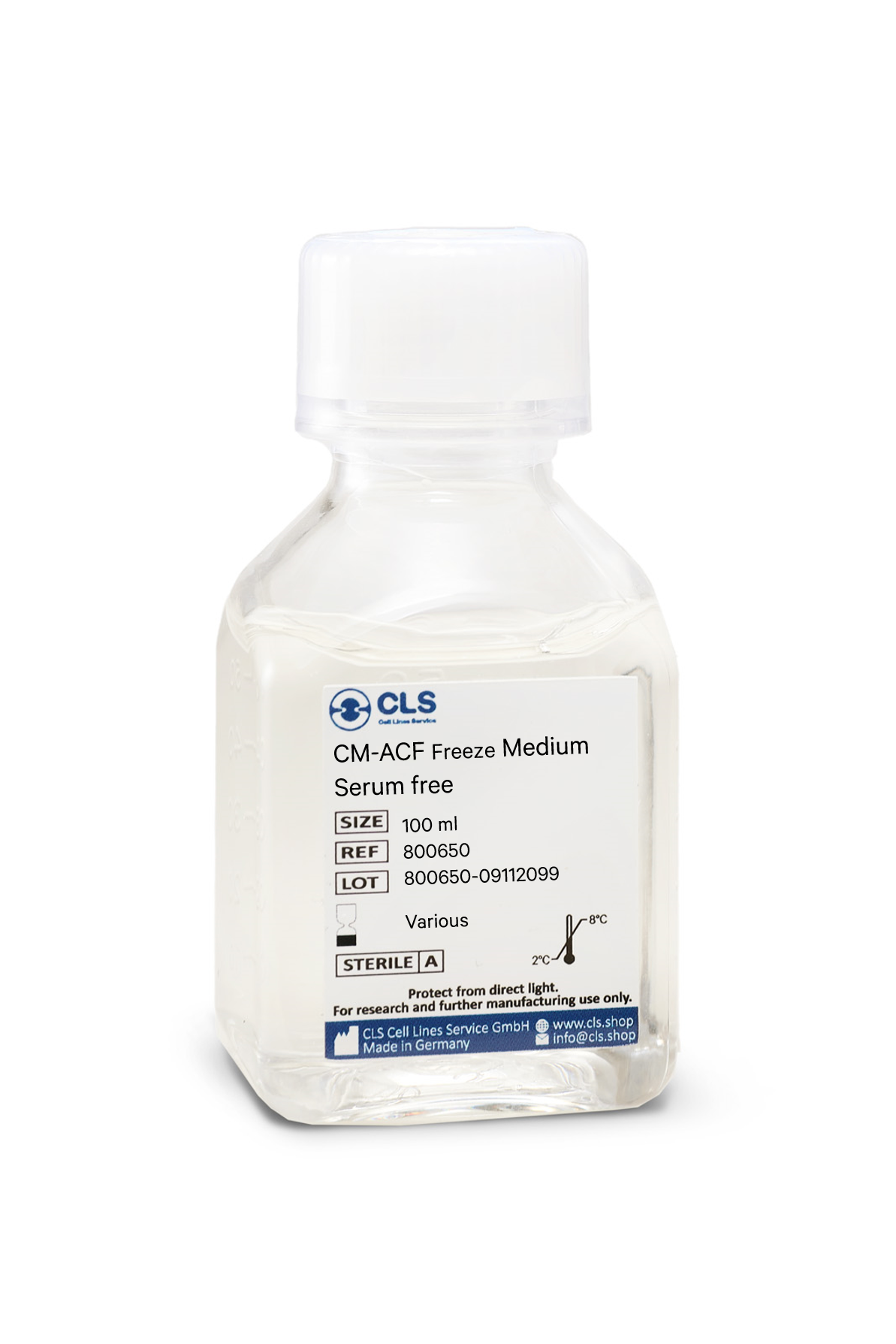
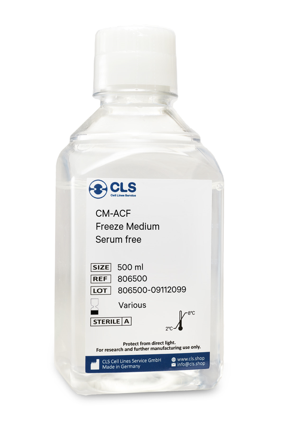
In biological research, the cryopreservation of mammalian cells is an invaluable tool. Successful preservation of cells is a top priority given that losing a cell line to contamination or improper storage conditions leads to lost time and money, ultimately delaying research results. Once the cells have been transferred from a cell growth medium to a freezing medium, the cells are typically frozen at a regulated rate and stored in liquid nitrogen vapor or at below -130°C in a mechanical deep freezer. The freeze medium CM-ACF enables cryopreservation of cells at below -130°C (or in liquid nitrogen), essentially eliminating the need for an additional, costly ultralow freezer and eliminating time-consuming and demanding controlled rate freezing processes. Simply collect the cells, aspirate the growth medium, resuspend in CM-ACF, transfer to a cryovial, and store the vial at below -130 °C.
Long shelf-life
CM-ACF is a serum-free, ready-to-use cryopreservation medium that can be stored in the refrigerator for up to one year.
Trusted by hundreds of researchers
Our advanced, serum-free cell freezing medium CM-ACF is a market-leading product in Germany and Europe and is distinguished by numerous publications involving hundreds of different cell lines worldwide. We tested it with more than 1000 cell lines from our proprietary cell bank.
Optimized serum-free ingredients
CM-ACF does not contain serum products. Serum-containing cryopreservation mediums have the disadvantage of fluctuating recovery rates and unclear composition. Since the composition and concentration of proteins and other biological components vary from batch to batch in serum, the reproducibility of experiments with cells that were frozen in a serum-containing medium may be compromised. As each component of CM-ACF is carefully defined, you can rest assured that cells always recover identically.
Contains DMSO, glucose, salts
Buffering capacity pH = 7.2 to 7.6
Universal
- even for stem cell preservation
All common cell lines can be frozen and thawed to yield many viable cells. Compared to standard media, the rate of recovery of even the most delicate cells is significantly higher. Using CM-ACF, we store over 1000 different cell lines with outstanding success.
Applications & Validation
The cells preserved in our CM-ACF freeze medium can be used for cell counting, viability and cryopreservation, cell culture, mammalian cell culture, gene expression analysis and genotyping, in vitro transcription, and polymerase chain reactions. Each batch's efficacy is evaluated using CHO-K1 cells. Each batch is tested for pH, osmolality, sterility, and endotoxins to ensure high quality.

