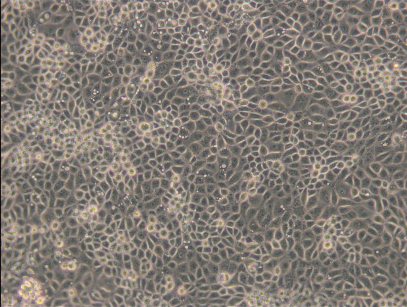
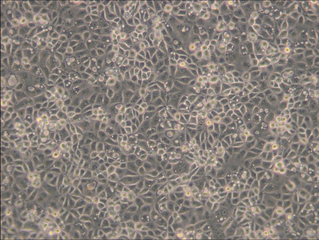
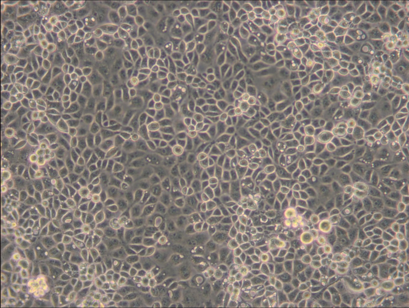
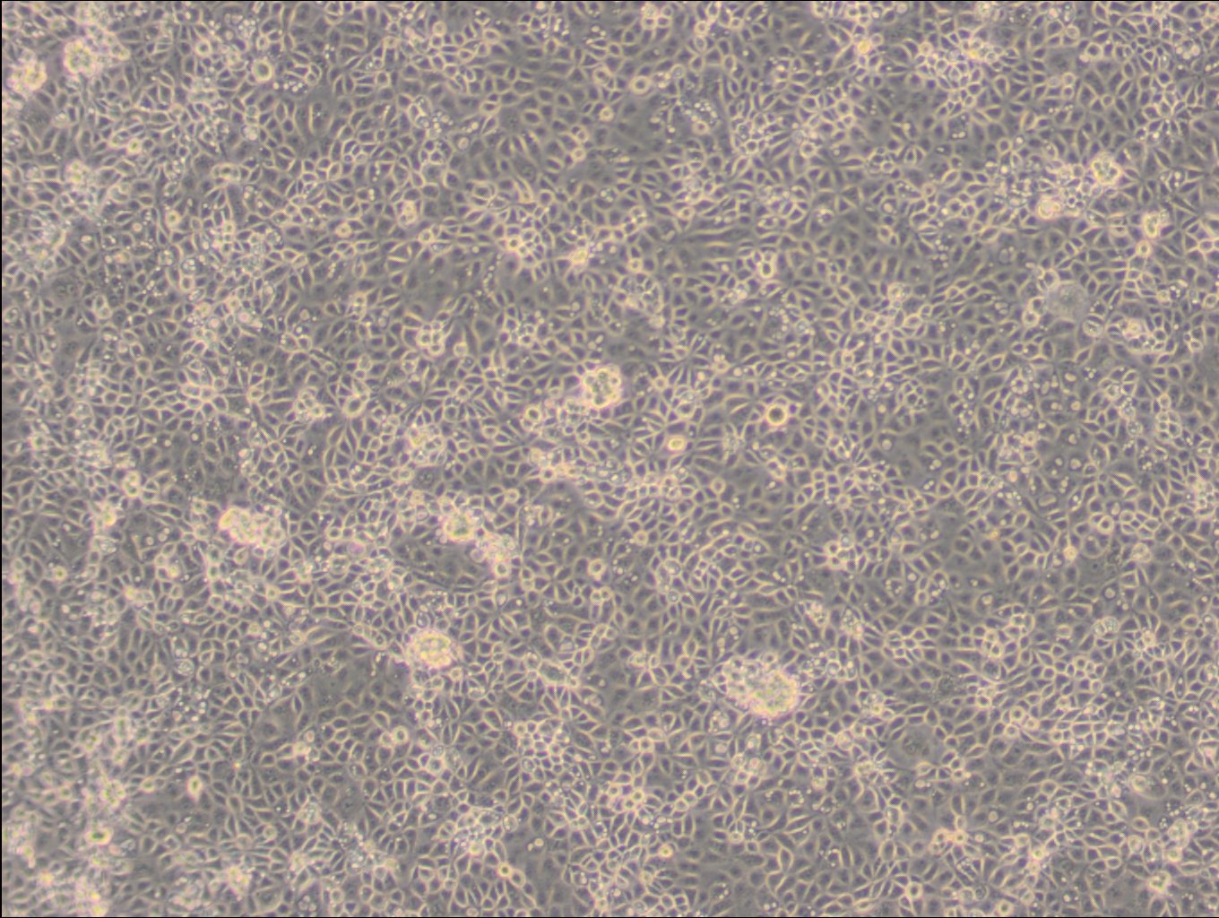
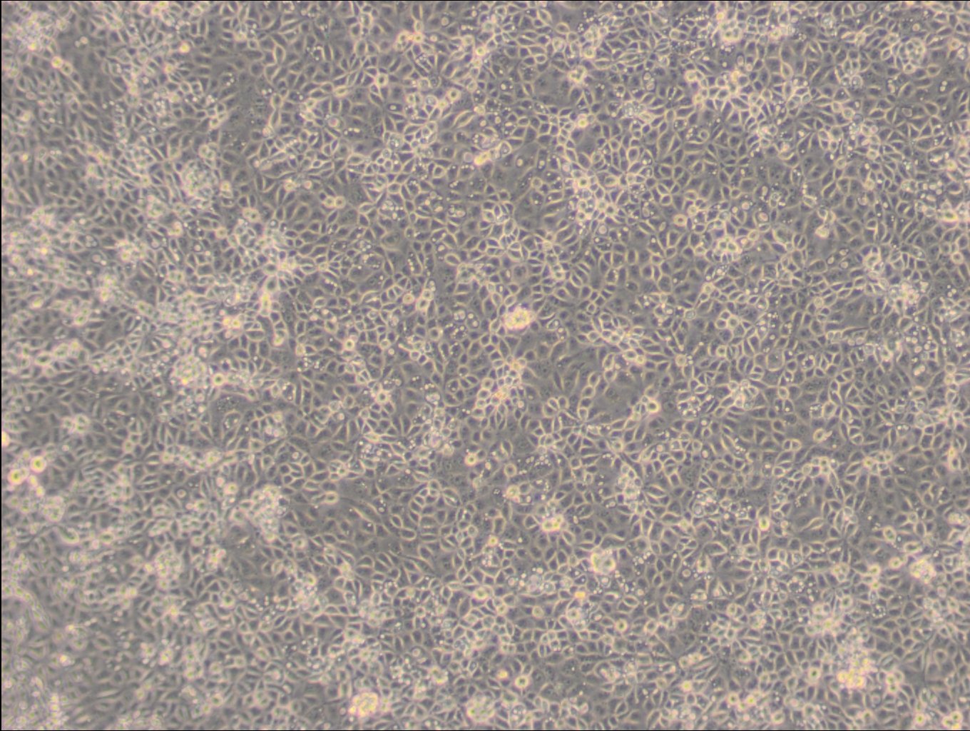
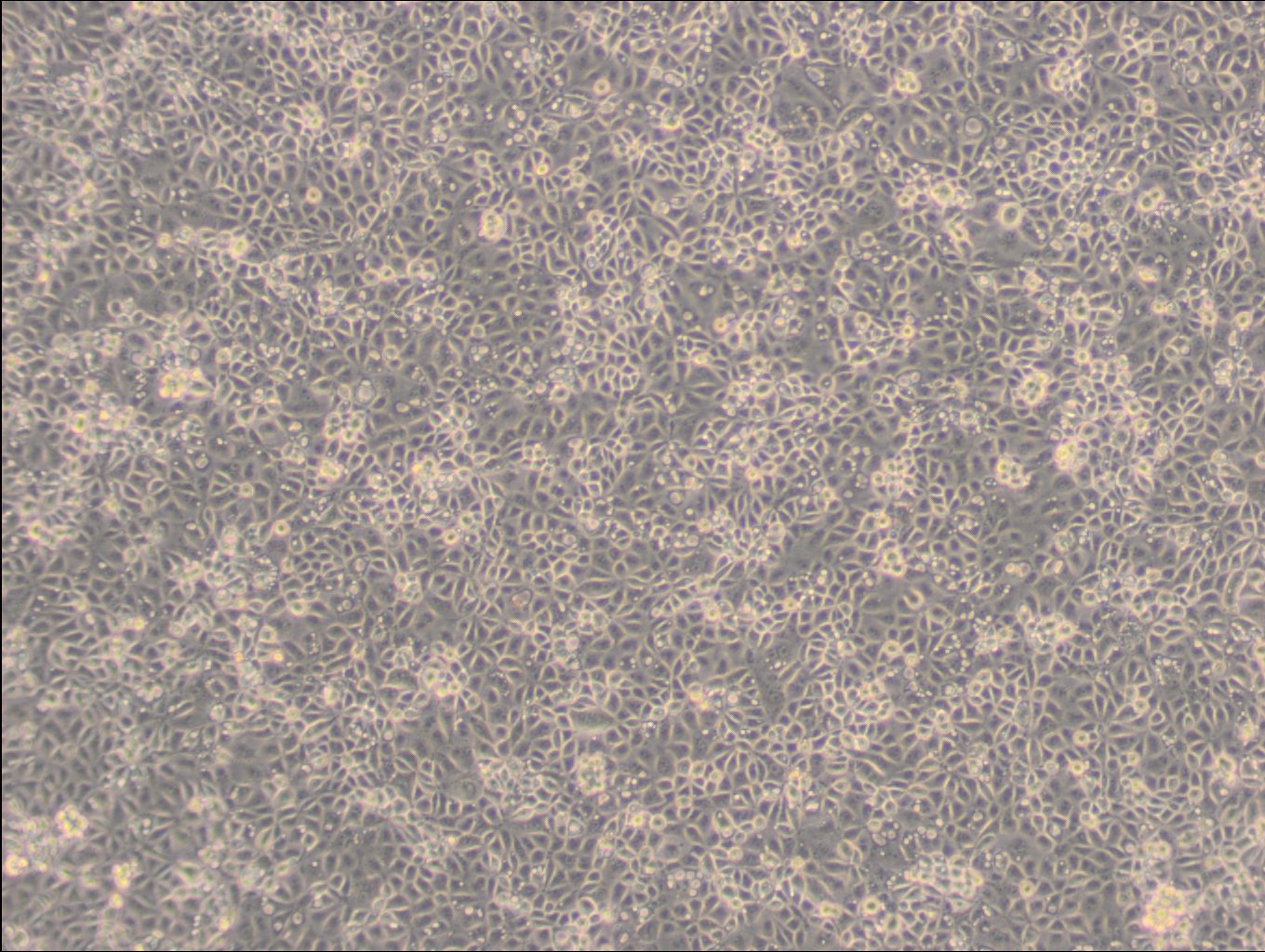
ST Cells
General information
Description | The ST cell line, derived from the connective tissue of a male Landrace pig, is primarily used in scientific studies related to virology and toxicology. These cells are of swine origin and are particularly valuable for research in veterinary medicine and comparative cellular biology, particularly for studies on viruses that affect swine. The fibroblast-like morphology of ST cells makes them a suitable model for studying cellular processes and virus-cell interactions in a porcine context. ST cells exhibit robust growth characteristics under standard cell culture conditions and have been utilized extensively to study a variety of swine pathogens, including foot-and-mouth disease virus and other Picornaviridae family members. Their susceptibility to different viral infections facilitates the analysis of viral life cycles, host-pathogen interactions, and the efficacy of antiviral compounds. Additionally, these cells are often used in the assessment of toxicological responses to various chemical agents, providing essential data on cellular responses and cytotoxicity in a non-human mammalian system. The versatility of the ST cell line in virological and toxicological assays underscores its utility in both fundamental and applied biological research. As such, ST cells continue to be a critical resource for researchers aiming to advance veterinary health, understand zoonotic disease mechanisms, and develop therapeutic strategies for diseases affecting swine populations. |
---|---|
Organism | Pig |
Tissue | Testis |
Synonyms | Swine Testis, STOMA24, Stoma 24, ST-IOWA |
Characteristics
Age | 80 to 90 days gestation |
---|---|
Gender | Male |
Morphology | Fibroblast |
Growth properties | Adherent |
Identifiers / Biosafety / Citation
Citation | ST (Cytion catalog number 305214) |
---|---|
Biosafety level | Biosafety level 1. The cell line harbors Porcine type-C oncovirus (PCOV) sequences and their transcripts, and the possibility of viral secretion cannot be excluded. In Germany, these viruses are categorized as BSL 1 for humans and BSL 2 for animals (TRBA 462). However, the German Central Committee on Biological Safety (ZKBS) assigns a BSL 2 classification to these viruses and infected cell lines when used for genetic modification purposes. |
Expression / Mutation
Handling
Culture Medium | EMEM, w: 2 mM L-Glutamine, w: 1.5 g/L NaHCO3, w: EBSS, w: 1 mM Sodium pyruvate, w: NEAA (Cytion article number 820100c) |
---|---|
Medium supplements | Supplement the medium with 10% FBS |
Passaging solution | Accutase |
Subculturing | Remove the old medium from the adherent cells and wash them with PBS that lacks calcium and magnesium. For T25 flasks, use 3-5 ml of PBS, and for T75 flasks, use 5-10 ml. Then, cover the cells completely with Accutase, using 1-2 ml for T25 flasks and 2.5 ml for T75 flasks. Let the cells incubate at room temperature for 8-10 minutes to detach them. After incubation, gently mix the cells with 10 ml of medium to resuspend them, then centrifuge at 300xg for 3 minutes. Discard the supernatant, resuspend the cells in fresh medium, and transfer them into new flasks that already contain fresh medium. |
Split ratio | 1:2 to 1:4 |
Fluid renewal | 2 to 3 times per week |
Freeze medium | CM-1 (Cytion catalog number 800100) |
Handling of cryopreserved cultures |
|
Quality control / Genetic profile / HLA
Sterility | Mycoplasma contamination is excluded using both PCR-based assays and luminescence-based mycoplasma detection methods. To ensure there is no bacterial, fungal, or yeast contamination, cell cultures are subjected to daily visual inspections. |
---|