HMEC-1 Cells


General information
| Description | HMEC-1 cells, or Human Microvascular Endothelial Cells-1, are an immortalized cell line derived from human dermal microvascular endothelial cells. This cell line was developed to facilitate research on the microvascular endothelial function and pathology. HMEC-1 cells are extensively used in vascular biology research due to their ability to retain many of the phenotypic and functional characteristics of primary endothelial cells. HMEC-1 cells display typical endothelial cell markers such as CD31 (PECAM-1), von Willebrand factor, and VE-cadherin, and they can form capillary-like structures when cultured on appropriate matrices, mimicking angiogenesis in vitro. This makes them particularly valuable for studies on angiogenesis, the formation of new blood vessels from pre-existing vasculature, a critical process in both physiological and pathological conditions such as wound healing, cancer growth, and cardiovascular diseases. These cells are also used to explore endothelial cell responses to inflammatory cytokines, the barrier function of endothelial layers, and the interaction between endothelial cells and other cell types like immune cells. HMEC-1 cells are amenable to genetic manipulation, allowing researchers to investigate the impact of specific genes on endothelial function and to model various vascular diseases. Furthermore, HMEC-1 cells serve as a model system for studying the permeability of endothelial barriers, which is crucial in the context of drug delivery and the pathogenesis of infectious diseases where pathogens cross endothelial barriers. The cell line?s versatility and ease of use continue to make it a cornerstone in studies of microvascular endothelial cell biology and pathology. |
|---|---|
| Organism | Human |
| Tissue | Skin |
| Applications | Research studies for human dermal endothelial cells |
| Synonyms | Hmec-1, HMEC1, CDC/EU.HMEC-1, Human Microvascular Endothelial Cell line-1 |
Characteristics
| Age | 1 month |
|---|---|
| Gender | Male |
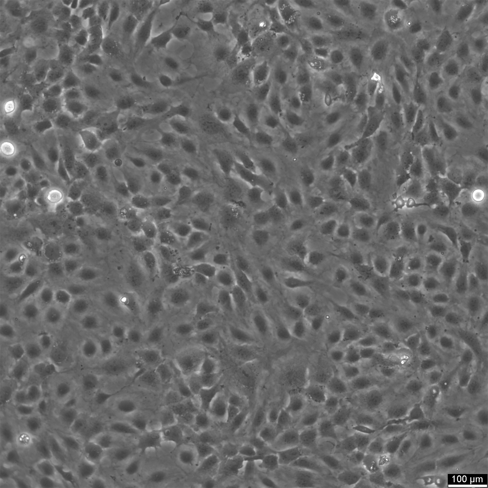
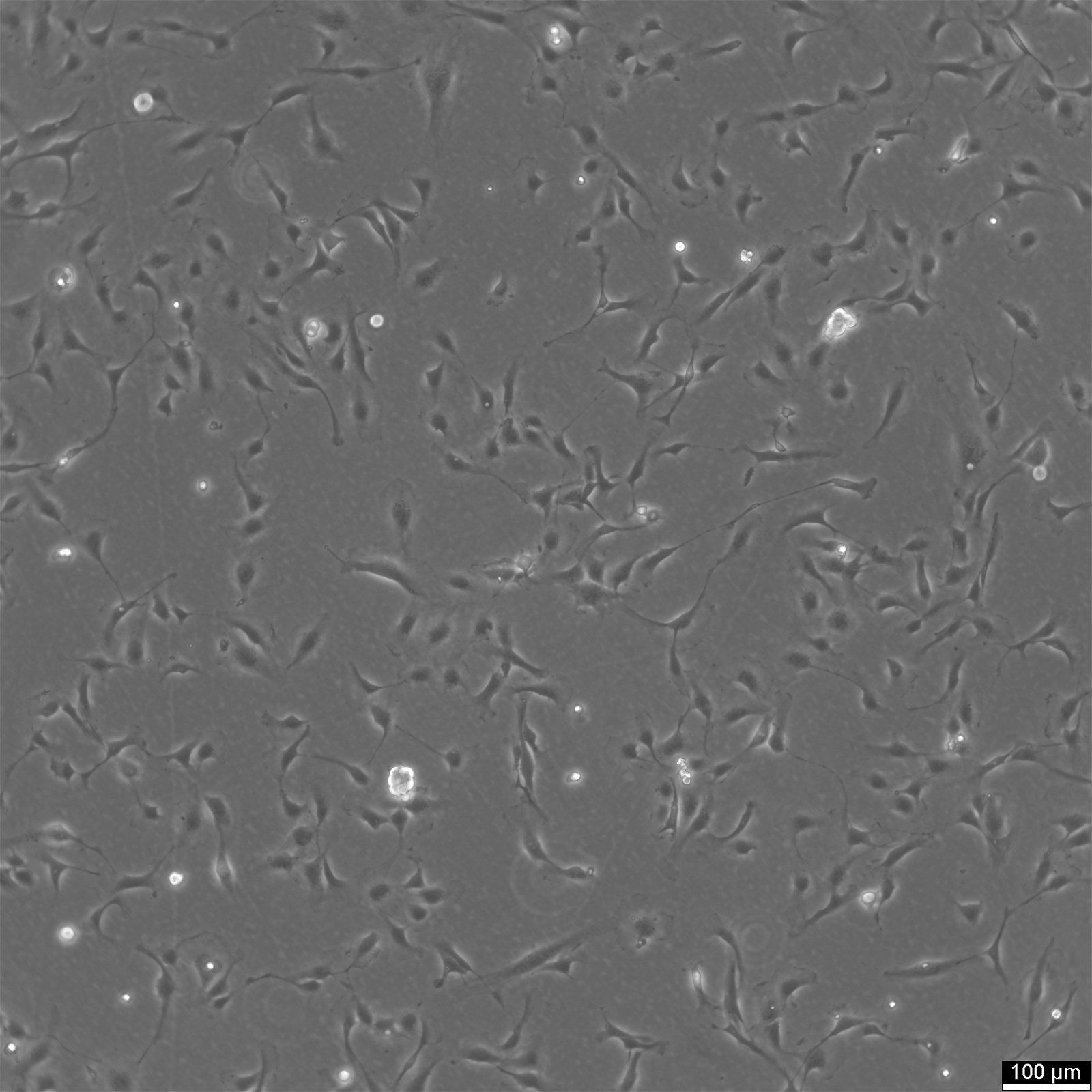
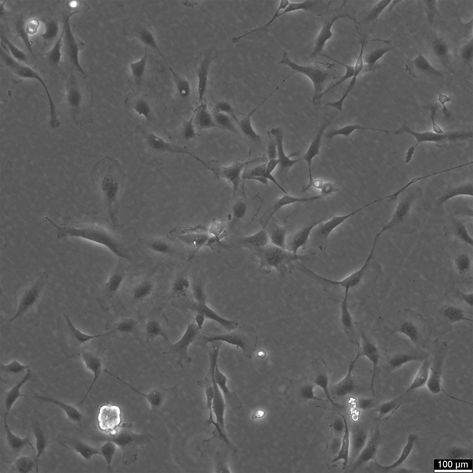
| Morphology | Endothelial-like |
| Growth properties | Adherent |
Identifiers / Biosafety / Citation
| Citation | HMEC-1 (Cytion catalog number 304064) |
|---|
Expression / Mutation
| Protein expression | von Willebrand's factor (vWF), cell adhesion molecules ICAM-1 |
|---|---|
| Viruses | Simian virus 40 (large T antigen) |
Handling
| Culture Medium | MCDB131 (We do not supply this product; please consider other suppliers. Please let us know if you need further assistance) |
|---|---|
| Medium supplements | Supplement the medium with 10% FBS, 3.0 g/L glucose, 2.2 g/L NaHCO3, 10 ng/mL Epidermal Growth Factor, 1 microgram/mL Hydrocortisone, 10 mM Glutamine |
| Passaging solution | Accutase |
| Subculturing | Remove the old medium from the adherent cells and wash them with PBS that lacks calcium and magnesium. For T25 flasks, use 3-5 ml of PBS, and for T75 flasks, use 5-10 ml. Then, cover the cells completely with Accutase, using 1-2 ml for T25 flasks and 2.5 ml for T75 flasks. Let the cells incubate at room temperature for 8-10 minutes to detach them. After incubation, gently mix the cells with 10 ml of medium to resuspend them, then centrifuge at 300xg for 3 minutes. Discard the supernatant, resuspend the cells in fresh medium, and transfer them into new flasks that already contain fresh medium. |
| Split ratio | 1:6 to 1:12 |
| Freeze medium | CM-1 (Cytion catalog number 800100) or CM-ACF (Cytion catalog number 806100) |
| Handling of cryopreserved cultures |
|
Quality control / Genetic profile / HLA
| Sterility | Mycoplasma contamination is excluded using both PCR-based assays and luminescence-based mycoplasma detection methods. To ensure there is no bacterial, fungal, or yeast contamination, cell cultures are subjected to daily visual inspections. |
|---|
