HK-2 Cells


General information
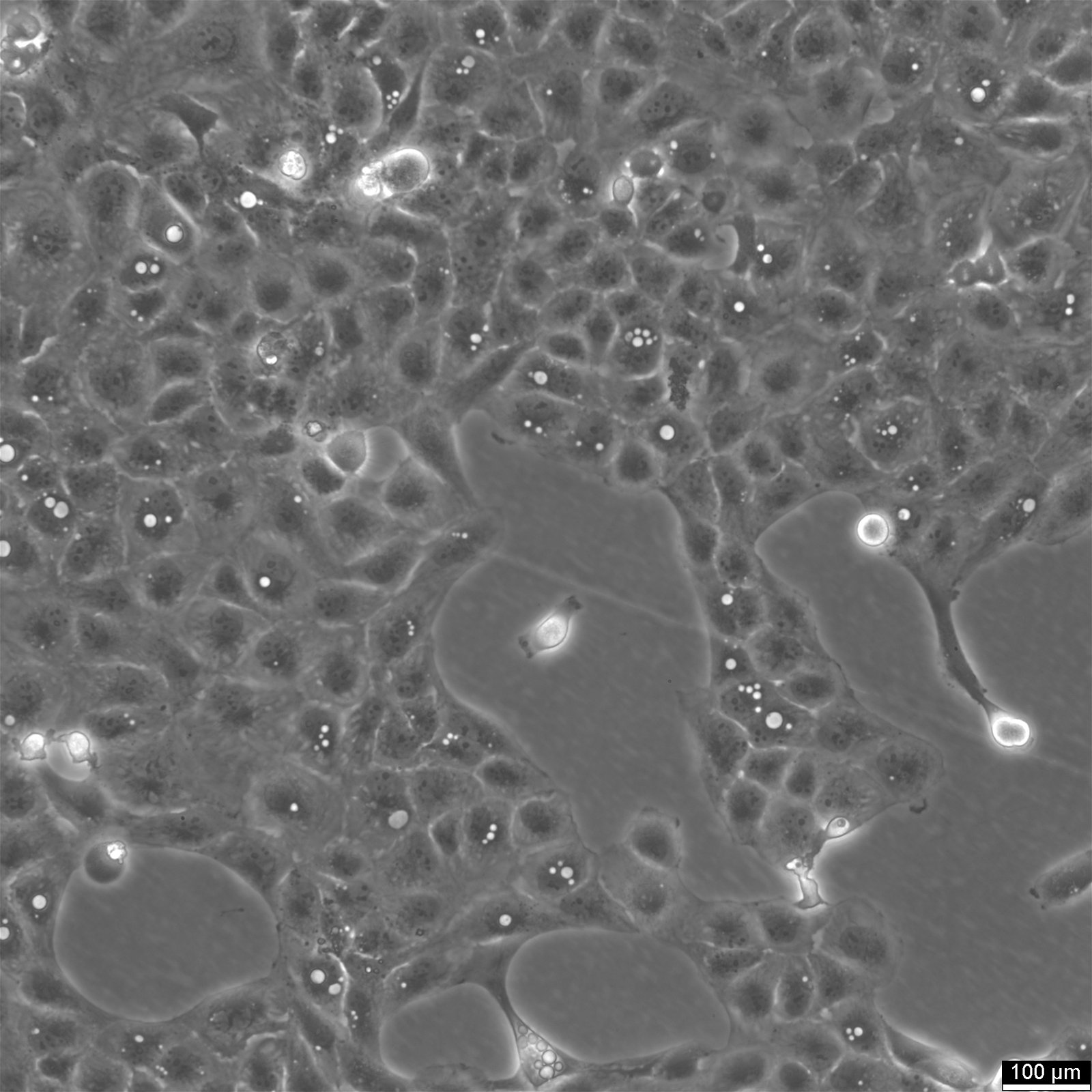
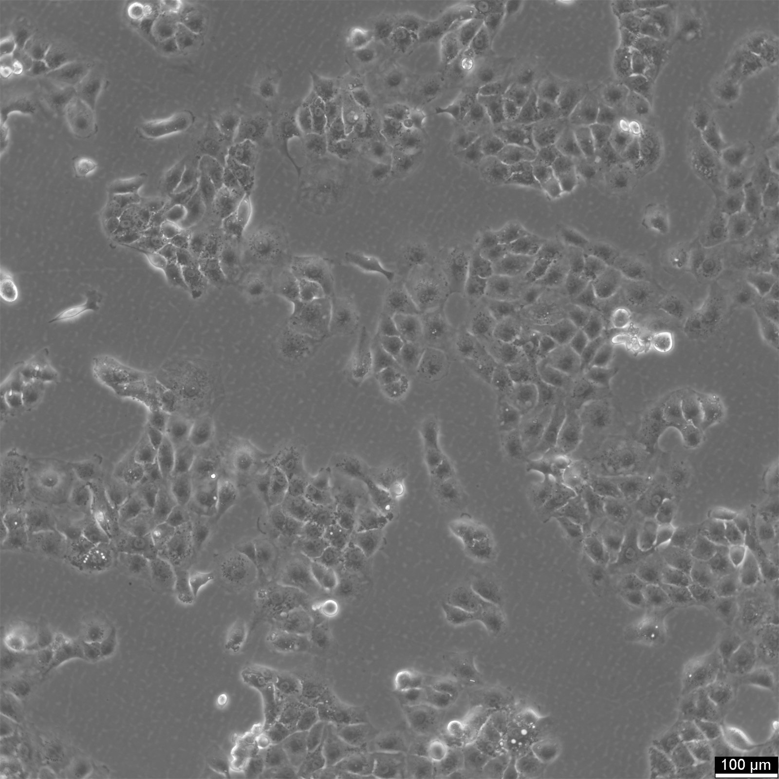
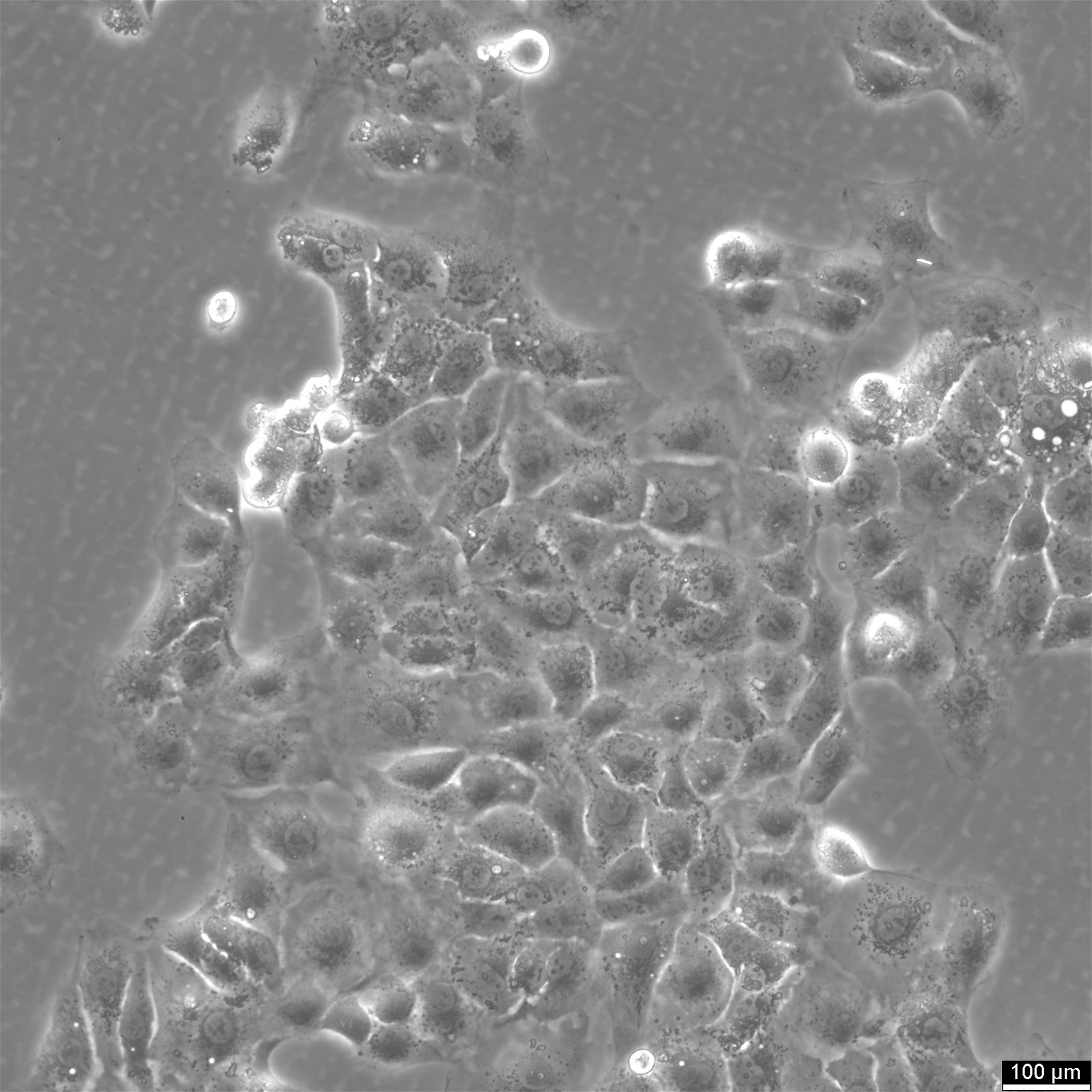
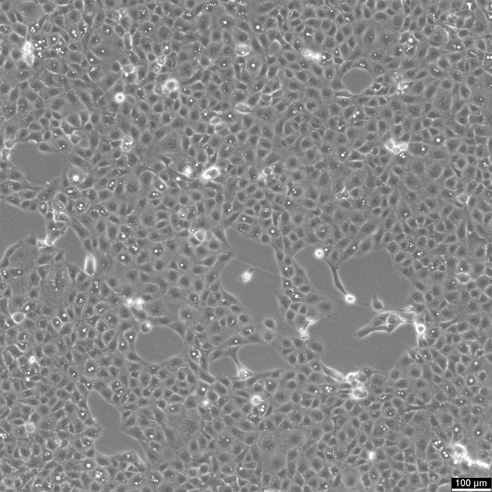
| Description | The human kidney-2 (HK-2) cell line is a type of proximal tubular cell (PTC) derived from a normal human kidney. HK-2 cells were created by transfection with a recombinant retrovirus containing the human papillomavirus 16 (HPV-16) E6/E7 genes. This process led to the immortalization of the cells and the establishment of a continuously growing cell line. The HK-2 cells have been extensively characterized, and research has shown that they retain a phenotype indicative of well-differentiated PTCs. The cells express EGF and require it for their growth and survival. They are positive for various genes, including alkaline phosphatase, gamma glutamyltranspeptidase, leucine aminopeptidase, acid phosphatase, cytokeratin, alpha 3, beta 1 integrin, and fibronectin. Additionally, the cells retain functional characteristics of proximal tubular and are capable of gluconeogenesis. HK-2 cells are anchorage-dependent, which means that they require a surface to adhere to in order to grow. They cannot grow in methylcellulose, soft agar, or suspension. The cells have a mean diameter of 18.2 micrometer, and their doubling time ranges between 47.3 h and 61.7 h. In terms of applications, HK-2 cells have been widely used in toxicology research due to their ability to reproduce experimental results obtained with freshly isolated PTCs. For example, HK-2 cells have been used to study the effects of environmental toxins, such as cadmium and cisplatin, on kidney cells. The cells have also been used to study the mechanisms underlying kidney diseases such as diabetic nephropathy and acute kidney injury. However, it is essential to note that the susceptibility of HK-2 cells to toxic compounds can be affected by the number of passages. In conclusion, HK-2 cells are a well-characterized PTC cell line that can be used in a variety of biological research applications, particularly in toxicology. However, researchers should be aware of the potential effects of passaging on the susceptibility of these cells to toxic compounds, and the number of passages should be considered when interpreting experimental results. |
|---|---|
| Organism | Human |
| Tissue | Kidney, cortex, proximal tubule |
| Synonyms | Hk-2, HK2, Human Kidney-2 |
Characteristics
| Age | Adult |
|---|---|
| Gender | Male |
| Ethnicity | European |
| Morphology | Epithelial |
| Growth properties | Adherent |
Identifiers / Biosafety / Citation
| Citation | HK-2 (Cytion catalog number 305021) |
|---|---|
| Biosafety level | 1 |
Expression / Mutation
| Receptors expressed | Epidermal growth factor(EGF), expressed |
|---|---|
| Protein expression | Alkaline Phosphatase, Gamma Glutamyltranspeptidase, Leucine Aminopeptidase, Acid Phosphatase, Cytokeratin, Alpha 3, Beta 1 Integrin, Fibronectin |
Handling
| Culture Medium | DMEM:Ham's F12, w: 3.1 g/L Glucose, w: 1.6 mM L-Glutamine, w: 15 mM HEPES, w: 1.0 mM Sodium pyruvate, w: 1.2 g/L NaHCO3 (Cytion article number 820400a) |
|---|---|
| Medium supplements | Supplement the medium with 10% FBS, 10 microgram/L IGF-1, 10 microgram/L EGF, 1 mg/L Transferrin, 0.5 microgram/L TGF-b1, 0,2 mg/L Biotin 0.05 mg/ml BPE |
| Passaging solution | Accutase |
| Subculturing | Remove the old medium from the adherent cells and wash them with PBS that lacks calcium and magnesium. For T25 flasks, use 3-5 ml of PBS, and for T75 flasks, use 5-10 ml. Then, cover the cells completely with Accutase, using 1-2 ml for T25 flasks and 2.5 ml for T75 flasks. Let the cells incubate at room temperature for 8-10 minutes to detach them. After incubation, gently mix the cells with 10 ml of medium to resuspend them, then centrifuge at 300xg for 3 minutes. Discard the supernatant, resuspend the cells in fresh medium, and transfer them into new flasks that already contain fresh medium. |
| Split ratio | 1:2 to 1:4 |
| Fluid renewal | 2 to 3 times per week |
| Freeze medium | CM-1 (Cytion catalog number 800100) or CM-ACF (Cytion catalog number 806100) |
| Handling of cryopreserved cultures |
|
Quality control / Genetic profile / HLA
| Sterility | Mycoplasma contamination is excluded using both PCR-based assays and luminescence-based mycoplasma detection methods. To ensure there is no bacterial, fungal, or yeast contamination, cell cultures are subjected to daily visual inspections. |
|---|---|
| STR profile |
Amelogenin: x,x
CSF1PO: 13
D13S317: 9
D16S539: 11,12
D5S818: 12
D7S820: 10,11
TH01: 9
TPOX: 8,9
vWA: 17,18
D3S1358: 16,17
D21S11: 28,30
D18S51: 12
Penta E: 10,11
Penta D: 9,12
D8S1179: 10,14
FGA: 20,22
D1S1656: 12,13
D6S1043: 12,13
D2S1338: 17,25
D12S391: 17.3,22
D19S433: 15,15.2
|
