HEp-2 Cells
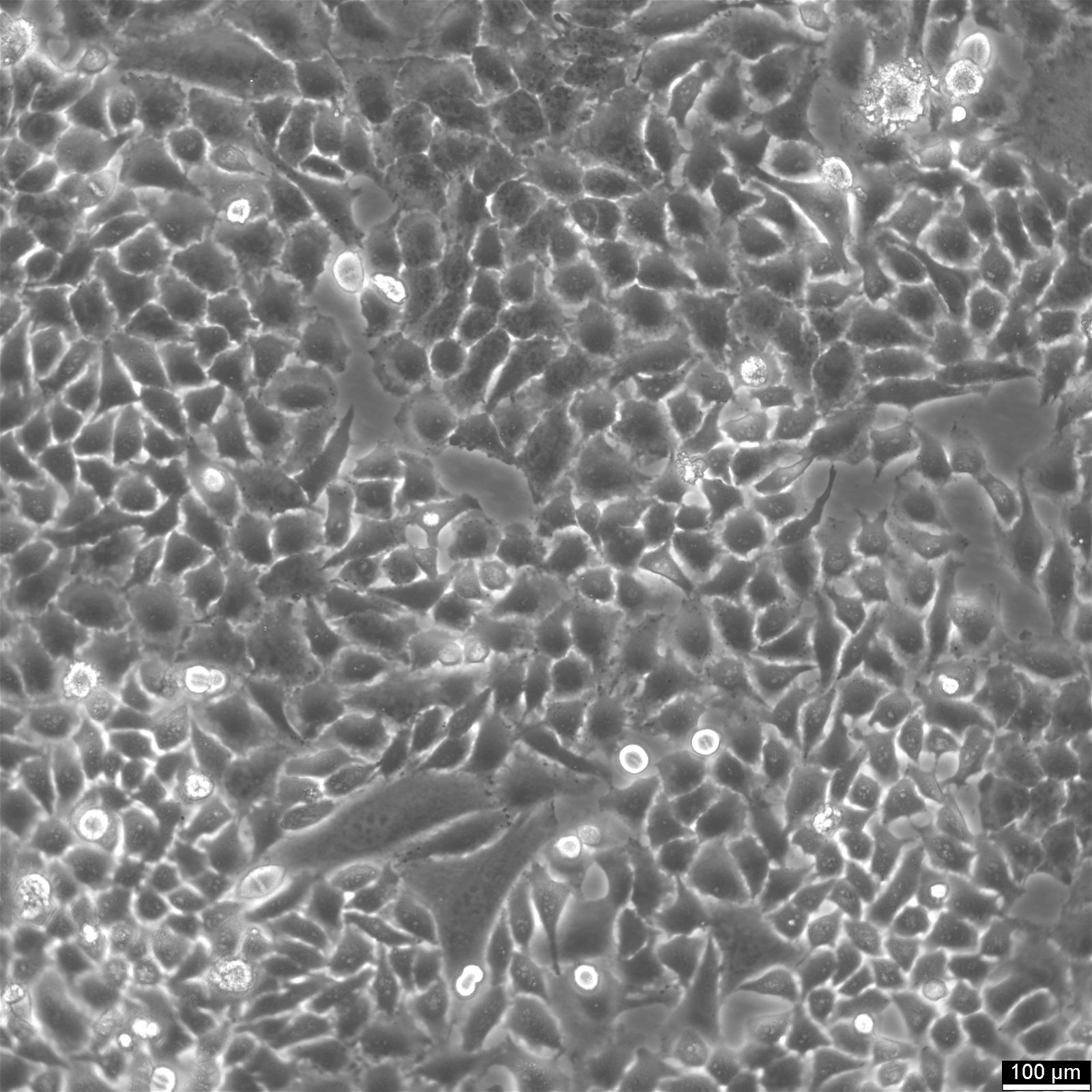
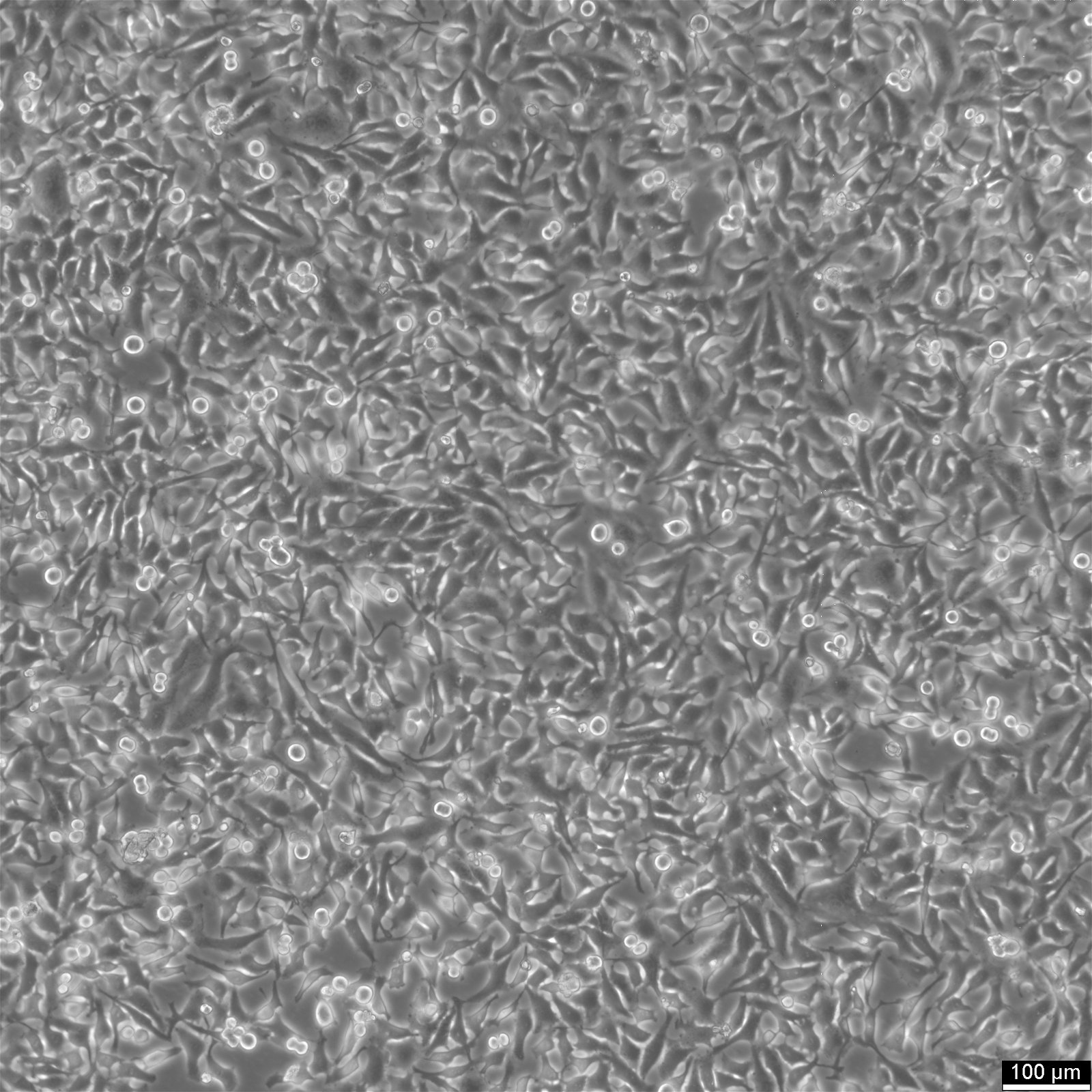
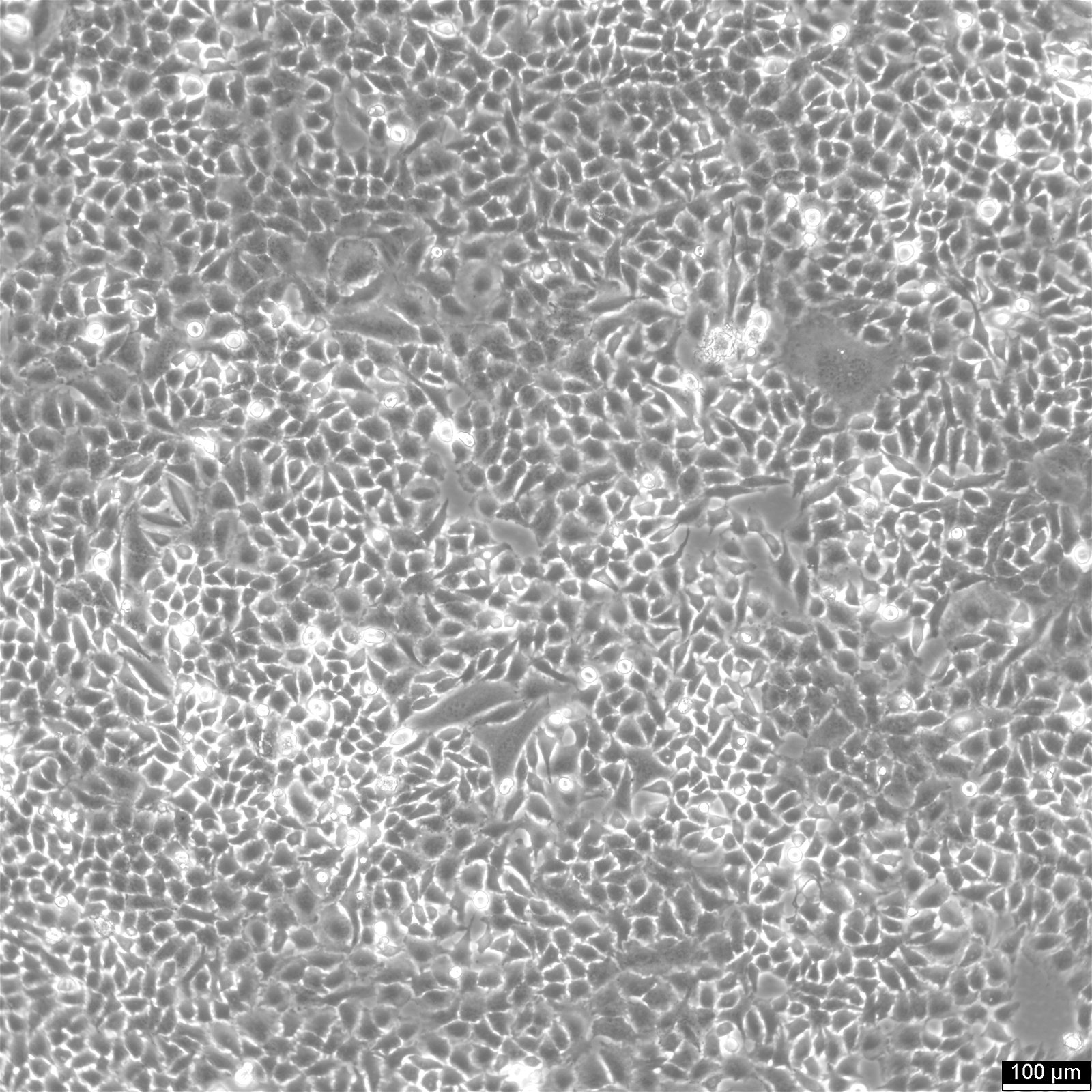


Introduction to Hep-2 cells
| Description | The HEp-2 cell line, originally believed to be derived from laryngeal cancer cells, was later identified through DNA fingerprinting and the presence of HeLa marker chromosomes as being contaminated with HeLa cells, a cell line that was derived from cervical cancer. Despite this, the HEp-2 cell line remains extensively utilized in indirect immunofluorescence to detect antinuclear antibodies (ANAs), which are key in diagnosing conditions like systemic lupus erythematosus and systemic sclerosis. The indirect immunofluorescence assay (IIFA) using HEp-2 cells, which provides clear positive or negative results, is the standard method for testing antinuclear antibodies. This straightforward approach is crucial for diagnosing and classifying different systemic autoimmune diseases. The patterns of autoantibodies observed in indirect immunofluorescence on HEp-2 cells, especially in the context of rheumatology, provide invaluable insights into various rheumatic diseases. Furthermore, the comprehensive review of antigens expressed by HEp-2 human cells under different culture conditions enables the identification of specific ANAs linked to diseases like lupus. In conclusion, while the contamination of cell lines like HEp-2 with HeLa cells has prompted concerns in cancer research about the accuracy and reliability of results and their clinical relevance, the utility of Hep-2 in the detection of antinuclear antibodies and its application across various research disciplines underscore its continued importance. The HEp-2 cell line serves as an essential tool in diagnosing and classifying autoimmune diseases, among other applications. |
|---|---|
| Organism | Human |
| Tissue | Larynx |
| Disease | Adenocarcinoma |
| Applications | In rheumatology, indirect immunofluorescence using HEp-2 cells plays a crucial role in diagnosing autoimmune diseases, including systemic lupus erythematosus and systemic sclerosis |
| Synonyms | Hep-2, HEP-2, HEp-2/HeLa, Hep 2, Hep2, HEp2, HEP2, H.Ep.-2, H.Ep. #2, H.Ep. No. 2, Hep II, Human Epidermoid carcinoma #2, Human Epithelioma-2 |
Details of the HEp-2 cell line
| Age | 30 years |
|---|---|
| Gender | Female |
| Ethnicity | African American |
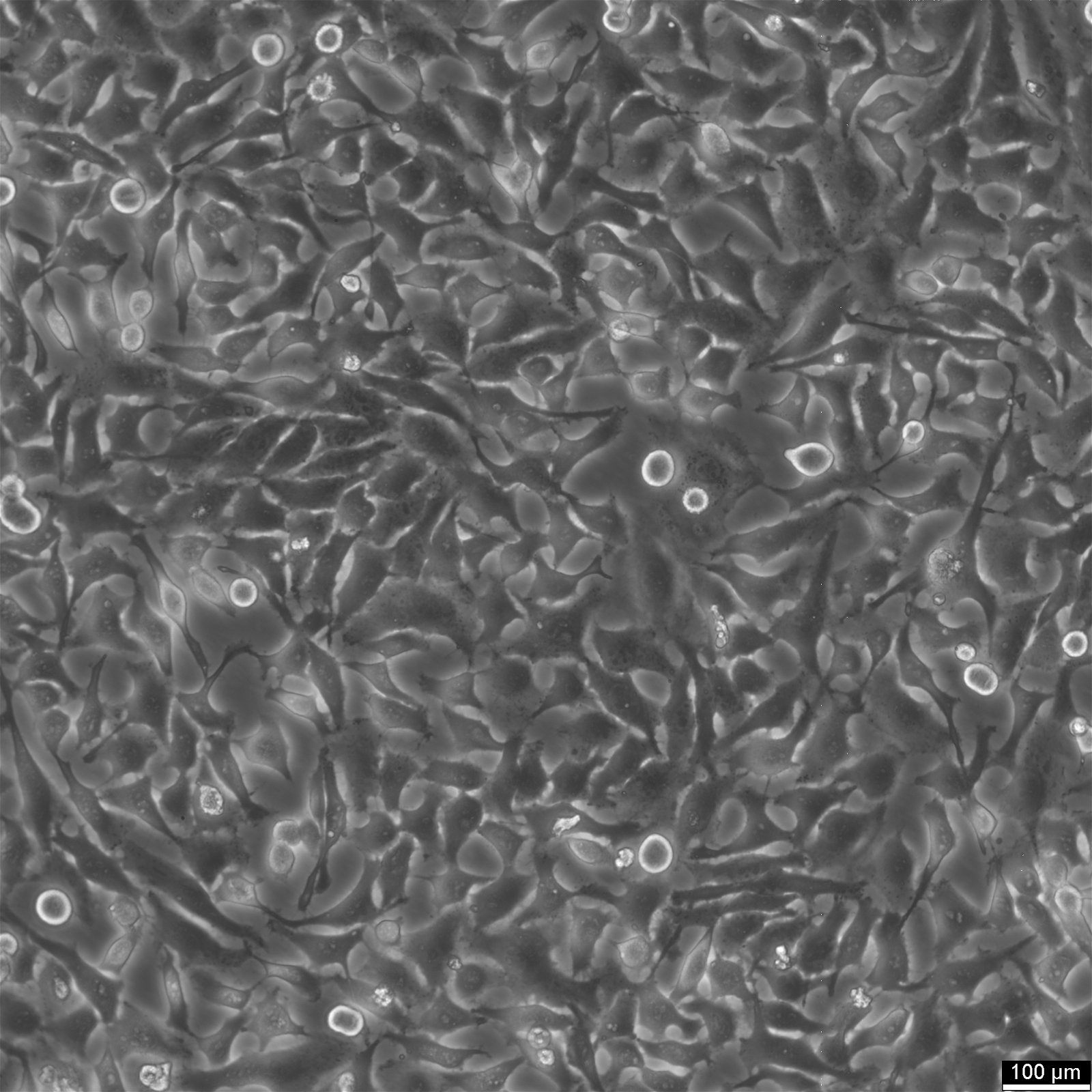
| Morphology | Epithelial-like |
| Growth properties | Monolayer, adherent |
Documentation
| Citation | HEp-2 (Cytion catalog number 300397) |
|---|---|
| Biosafety level | 1 |
Genetic profile
| Isoenzymes | G6PD, A |
|---|---|
| Reverse transcriptase | Negative |
| Products | Keratin |
Handling of Hep2 cells
| Culture Medium | EMEM, w: 2 mM L-Glutamine, w: 1.5 g/L NaHCO3, w: EBSS, w: 1 mM Sodium pyruvate, w: NEAA (Cytion article number 820100c) |
|---|---|
| Medium supplements | Supplement the medium with 10% FBS |
| Passaging solution | Accutase |
| Subculturing | Remove the old medium from the adherent cells and wash them with PBS that lacks calcium and magnesium. For T25 flasks, use 3-5 ml of PBS, and for T75 flasks, use 5-10 ml. Then, cover the cells completely with Accutase, using 1-2 ml for T25 flasks and 2.5 ml for T75 flasks. Let the cells incubate at room temperature for 8-10 minutes to detach them. After incubation, gently mix the cells with 10 ml of medium to resuspend them, then centrifuge at 300xg for 3 minutes. Discard the supernatant, resuspend the cells in fresh medium, and transfer them into new flasks that already contain fresh medium. |
| Split ratio | A ratio of 1:4 to 1:10 is recommended |
| Seeding density | 1 x 10^4 cells/cm^2 |
| Fluid renewal | 2 to 3 times per week |
| Freezing recovery | After thawing, plate the cells at 5 x 10^4 cells/cm^2 and allow the cells to recover from the freezing process and to adhere for at least 24 hours. |
| Freeze medium | CM-1 (Cytion catalog number 800100) or CM-ACF (Cytion catalog number 806100) |
| Handling of cryopreserved cultures |
|
Quality control
| Sterility | Mycoplasma contamination is excluded using both PCR-based assays and luminescence-based mycoplasma detection methods. To ensure there is no bacterial, fungal, or yeast contamination, cell cultures are subjected to daily visual inspections. |
|---|---|
| STR profile |
Amelogenin: x,x
CSF1PO: 9,10
D13S317: 12,13.3
D16S539: 9,10
D5S818: 11,12
D7S820: 8,12
TH01: 7
TPOX: 8,12
vWA: 16,18
D3S1358: 15,18
D21S11: 27,28
D18S51: 16
Penta E: 7,17
Penta D: 8,15
D8S1179: 12,13
FGA: 18,21
|
Required products
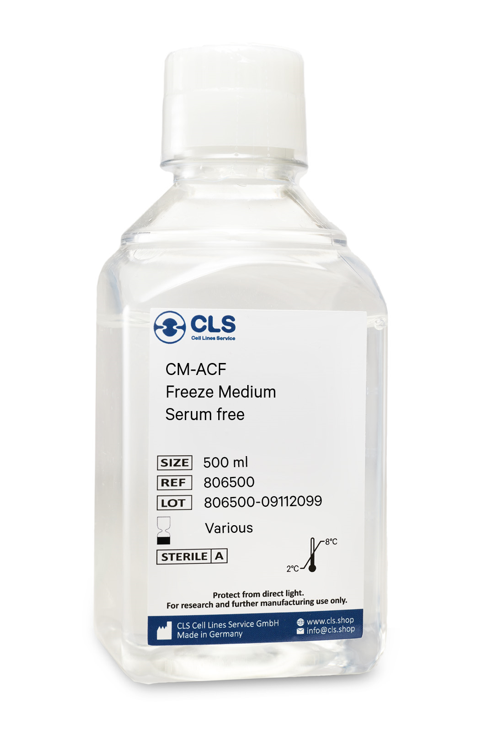
In biological research, the cryopreservation of mammalian cells is an invaluable tool. Successful preservation of cells is a top priority given that losing a cell line to contamination or improper storage conditions leads to lost time and money, ultimately delaying research results. Once the cells have been transferred from a cell growth medium to a freezing medium, the cells are typically frozen at a regulated rate and stored in liquid nitrogen vapor or at below -130°C in a mechanical deep freezer. The freeze medium CM-ACF enables cryopreservation of cells at below -130°C (or in liquid nitrogen), essentially eliminating the need for an additional, costly ultralow freezer and eliminating time-consuming and demanding controlled rate freezing processes. Simply collect the cells, aspirate the growth medium, resuspend in CM-ACF, transfer to a cryovial, and store the vial at below -130 °C.
Long shelf-life
CM-ACF is a serum-free, ready-to-use cryopreservation medium that can be stored in the refrigerator for up to one year.
Trusted by hundreds of researchers
Our advanced, serum-free cell freezing medium CM-ACF is a market-leading product in Germany and Europe and is distinguished by numerous publications involving hundreds of different cell lines worldwide. We tested it with more than 1000 cell lines from our proprietary cell bank.
Optimized serum-free ingredients
CM-ACF does not contain serum products. Serum-containing cryopreservation mediums have the disadvantage of fluctuating recovery rates and unclear composition. Since the composition and concentration of proteins and other biological components vary from batch to batch in serum, the reproducibility of experiments with cells that were frozen in a serum-containing medium may be compromised. As each component of CM-ACF is carefully defined, you can rest assured that cells always recover identically.
Contains DMSO, glucose, salts
Buffering capacity pH = 7.2 to 7.6
Universal
- even for stem cell preservation
All common cell lines can be frozen and thawed to yield many viable cells. Compared to standard media, the rate of recovery of even the most delicate cells is significantly higher. Using CM-ACF, we store over 1000 different cell lines with outstanding success.
Applications & Validation
The cells preserved in our CM-ACF freeze medium can be used for cell counting, viability and cryopreservation, cell culture, mammalian cell culture, gene expression analysis and genotyping, in vitro transcription, and polymerase chain reactions. Each batch's efficacy is evaluated using CHO-K1 cells. Each batch is tested for pH, osmolality, sterility, and endotoxins to ensure high quality.
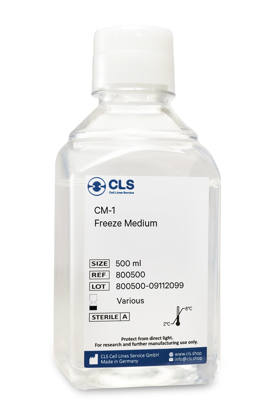
In biological research, the cryopreservation of mammalian cells is an invaluable tool. Successful preservation of cells is a top priority given that losing a cell line to contamination or improper storage conditions leads to lost time and money, ultimately delaying research results. Once the cells have been transferred from a cell growth medium to a freezing medium, the cells are typically frozen at a regulated rate and stored in liquid nitrogen vapor or at below -130°C in a mechanical deep freezer. The freeze medium CM-1 enables cryopreservation of cells at below -130°C (or in liquid nitrogen), essentially eliminating the need for an additional, costly ultralow freezer and eliminating time-consuming and demanding controlled rate freezing processes. Simply collect the cells, aspirate the growth medium, resuspend in CM-1, transfer to a cryovial, and store the vial at below -130 °C.
Long shelf-life
CM-1 is a serum-containing, ready-to-use cryopreservation medium that can be stored in the refrigerator for up to one year.
Trusted by hundreds of researchers
Our advanced cell freezing medium CM-1 is a market-leading product in Germany and Europe and is distinguished by numerous publications involving hundreds of different cell lines worldwide. We tested it with more than 1000 cell lines from our proprietary cell bank.
Optimized ingredients
CM-1 does contain serum products. Serum-containing cryopreservation mediums optimally protect the cells whilst being frozen and have the advantage of high recovery rates. As CM-1 has been tested with a multitude of cell lines, you can rest assured that your cells always recover well.
Contains FBS, DMSO, glucose, salts
Buffering capacity pH = 7.2 to 7.6
Applications & Validation
The cells preserved in our CM-1 freeze medium can be used for cell counting, viability and cryopreservation, cell culture, mammalian cell culture, gene expression analysis and genotyping, in vitro transcription, and polymerase chain reactions. Each batch's efficacy is evaluated using CHO-K1 cells. Each batch is tested for pH, osmolality, sterility, and endotoxins to ensure high quality.
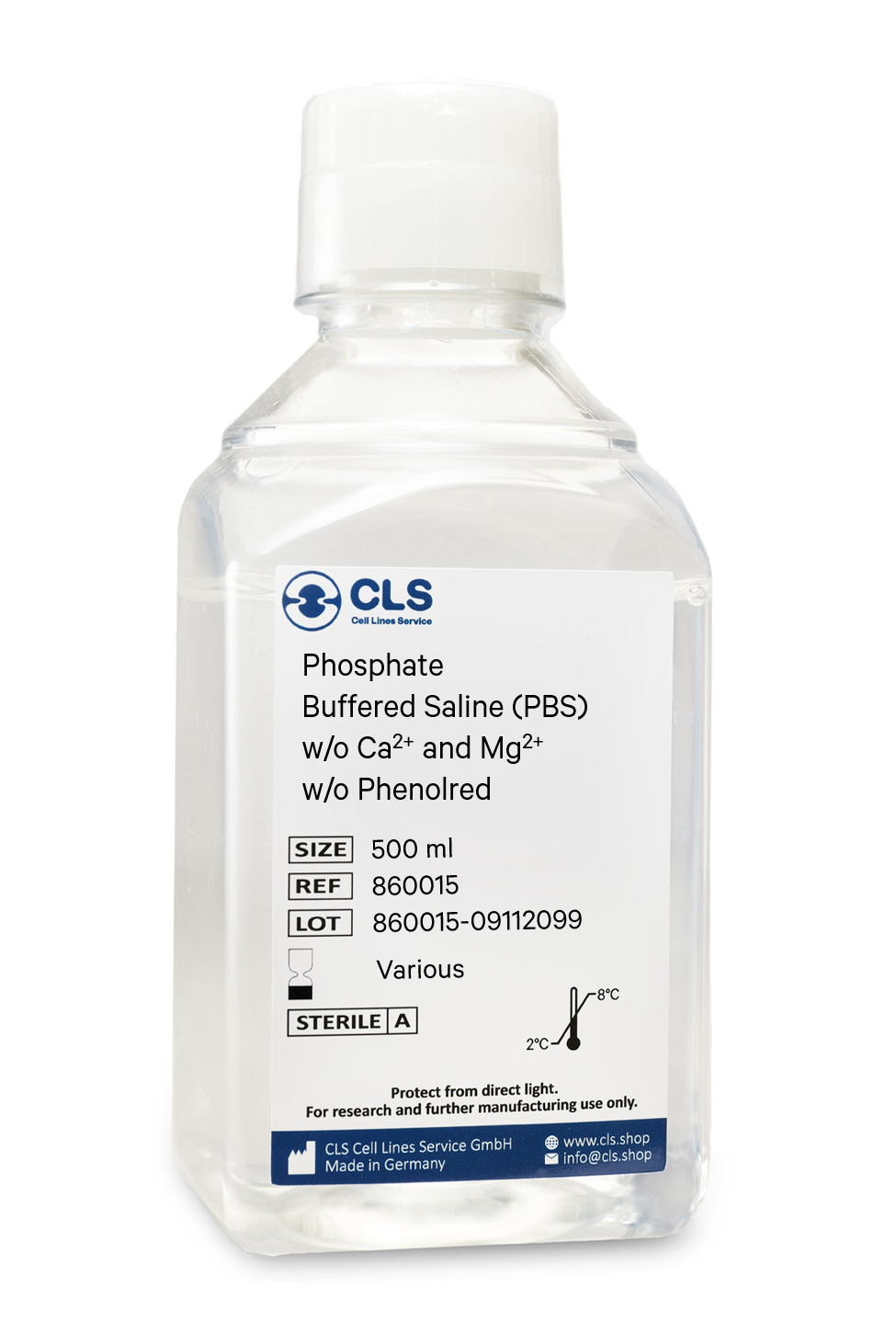
Phosphate-buffered saline (PBS) is a versatile buffer solution used in many biological and chemical applications, as well as tissue processing. Our PBS solution is formulated with high-quality ingredients to ensure a constant pH during experiments. The osmolarity and ion concentrations of our PBS solution are matched to those of the human body, making it isotonic and non-toxic to most cells.
Composition of our PBS Solution
Our PBS solution is a pH-adjusted blend of ultrapure-grade phosphate buffers and saline solutions. At a 1X working concentration, it contains 137 mM NaCl, 2.7 mM KCl, 8 mM Na2HPO4, and 2 mM KH2PO4. We have chosen this composition based on CSHL protocols and Molecular cloning by Sambrook, which are well-established standards in the research community.
Applications of our PBS Solution
Our PBS solution is ideal for a wide range of applications in biological research. Its isotonic and non-toxic properties make it perfect for substance dilution and cell container rinsing. Our PBS solution with EDTA can also be used to disengage attached and clumped cells. However, it is important to note that divalent metals such as zinc cannot be added to PBS as this may result in precipitation. In such cases, Good's buffers are recommended. Moreover, our PBS solution has been shown to be an acceptable alternative to viral transport medium for the transport and storage of RNA viruses, such as SARS-CoV-2.
Storage of our PBS Solution
Our PBS solution can be stored at room temperature, making it easy to use and access.
To sum up
In summary, our PBS solution is an essential component in many biological and chemical experiments. Its isotonic and non-toxic properties make it suitable for numerous applications, from cell culture to viral transport medium. By choosing our high-quality PBS solution, researchers can optimize their experiments and ensure accurate and reliable results.
Composition
Components
mg/L
Inorganic Salts
Potassium chloride
200,00
Potassium dihydrogen phosphate
200,00
Sodium chloride
8,000.00
di-Sodium hydrogen phosphate anhydrous
1,150.00

