
Published: 2023 | Last reviewed: May 2026
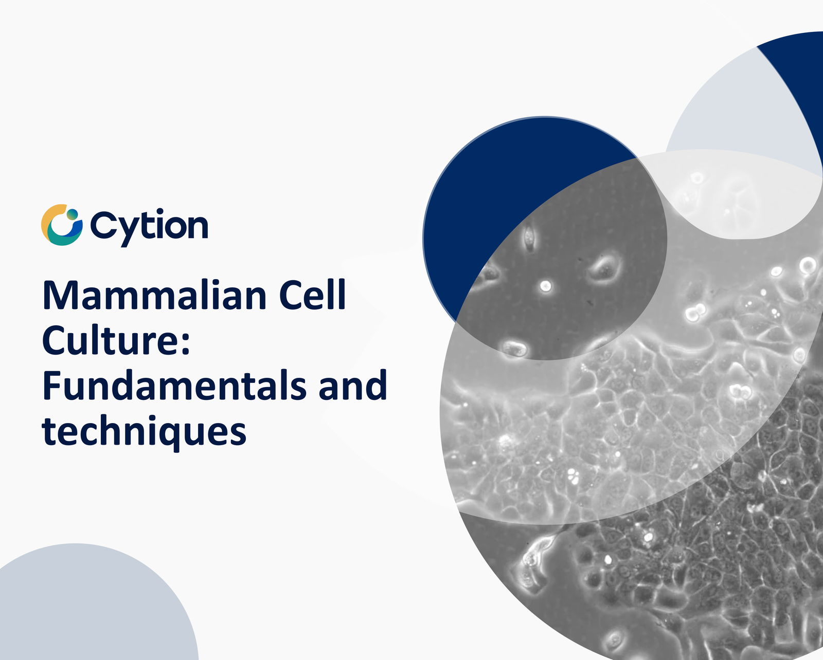
Mammalian Cell Culture: Fundamentals and Techniques
Mammalian cell culture is a cornerstone technique in biological research, allowing scientists to study cells in a controlled environment outside of living organisms. This process involves isolating cells from tissues, maintaining them in carefully controlled conditions, and propagating them for various experimental purposes. Mammalian cell culture is crucial for understanding cellular processes, disease mechanisms, and developing new therapies, including those using immortal cell lines.
Key Takeaways:
- Cells can be isolated from tissues using enzymatic digestion or explant culture methods
- Primary cells have limited lifespan, while immortalized cell lines can proliferate indefinitely
- Culture conditions, including growth media composition, are critical for cell survival and proliferation
- Cells can be grown in suspension or as adherent cultures, depending on their type and research needs
- Common culture media include MEM, DMEM, and RPMI 1640, each tailored for specific cell types
- Typical growth conditions involve 37°C temperature, 5% CO2, and 95% relative humidity
- Serum alternatives like human platelet lysate (hPL) are increasingly used to avoid potential contamination issues
Key Aspects of Mammalian Cell Culture
Cell Isolation
- Culture Media
Incubation Conditions
- Cell Analysis
Cell Isolation Techniques
The process of establishing a cell culture begins with isolating cells from tissues. There are several methods to accomplish this, each suited to different tissue types and research goals. For blood samples, cell isolation is relatively straightforward, with white blood cells being the primary focus for culture due to their growth capabilities. Solid tissues require more complex isolation techniques. One common method involves enzymatic digestion, where enzymes like collagenase, trypsin, or pronase are used to break down the extracellular matrix, releasing individual cells into suspension. Alternatively, researchers may employ the explant culture method, where small pieces of tissue are placed directly into growth media, allowing cells to migrate out and proliferate. The choice between these methods often depends on the specific tissue type, the desired cell population, and the intended experimental use. It's important to note that cells isolated directly from an organism are termed primary cells and, with some exceptions like tumor-derived cells, typically have a limited lifespan in culture before undergoing senescence.
Essential Products for Mammalian Cell Culture
| Product Name | Product Number | Category | Application |
|---|---|---|---|
| DMEM, w: 4.5 g/L Glucose, w: 4 mM L-Glutamine, w: 1.5 g/L NaHCO3, w: 1.0 mM Sodium pyruvate | 820300a | Culture Media | General purpose medium for various mammalian cell types |
| DMEM:Ham's F12 (1:1), w: 3.1 g/L Glucose, w: 1.6 mM L-Glutamine, w: 15 mM HEPES, w: 1.0 mM Sodium pyruvate, w: 1.2 g/L NaHCO3 | 820400a | Culture Media | Suitable for a wide range of mammalian cells, especially epithelial cells |
| RPMI 1640, w: 2.1 mM stable Glutamine, w: 2.0 g/L NaHCO3 | 820700a | Culture Media | Commonly used for lymphoid cells and hybrid cell lines |
| Accutase | 830100 | Cell Dissociation | Gentle cell detachment solution for adherent cells |
| Freeze Medium CM-1 | 800150 | Cryopreservation | For freezing and long-term storage of mammalian cells |
| Freeze medium CM-ACF, serum free | 800650 | Cryopreservation | Animal component-free medium for cell freezing |
| PBS | 860015 | Buffer Solution | For washing cells and maintaining pH balance |
| Endothelial Cell Growth Medium | 820731 | Specialized Media | Optimized for culture of endothelial cells |
| Mycoplasma testing | 900159 | Quality Control | Essential for detecting mycoplasma contamination in cultures |
| Cell line authentication - Human | 900154 | Quality Control | Verifies the identity of human cell lines |
This table represents a selection of essential products for mammalian cell culture. For our complete range of cell culture products, including specialized media and reagents, please visit our Media and Reagents category page.

Accutase is a ready-to-use, sterile-filtered cell detachment solution designed as a gentle alternative to trypsin/EDTA for dissociating adherent mammalian cells from standard tissue culture plasticware and adhesion-coated surfaces. It combines proteolytic and collagenolytic enzyme activity in a balanced salt solution to deliver effective yet controlled dissociation, preserving cell-surface proteins and supporting high post-passage viability and rapid reattachment.
The Accutase formulation is based on Dulbecco’s phosphate-buffered saline (DPBS) with EDTA and phenol red as a visual pH indicator. The enzymes are of non-mammalian and non-bacterial origin, making Accutase particularly well suited to stem cell research, vaccine workflows, and any application where animal
- or microbially-derived contaminants must be minimised. The solution auto-inhibits at 37 °C, so no neutralising reagent or serum-containing medium is required after detachment – cells can be transferred directly into fresh medium.
Key Features
Ready-to-use 1x sterile-filtered liquid – no dilution or reconstitution required
Combined proteolytic and collagenolytic enzyme activity for gentle dissociation
Each batch standardised to a defined dissociation activity for lot-to-lot consistency
Non-mammalian and non-bacterial enzyme origin
Auto-inhibits at 37 °C – no neutralising solution needed
Formulated in Dulbecco’s PBS with EDTA
Phenol red included as visual pH indicator
pH 6.8 – 7.8
Typical Applications
Accutase gently dissociates a wide variety of adherent and sensitive cell types, including human embryonic stem cells (hESCs), human induced pluripotent stem cells (iPSCs), neural stem cells, primary neurons, and routinely cultured adherent lines such as HeLa, HEK 293, CHO, MDCK, Vero, NIH/3T3, BHK-21 and A549. Typical use cases include:
Routine subculture and passaging of adherent mammalian cells
Gentle single-cell dissociation of hESCs, iPSCs and other sensitive lines
Sample preparation for flow cytometry and FACS analysis
Analysis of cell-surface markers where epitope integrity matters
Cell migration, proliferation and apoptosis assays
Quiescence assays by serum starvation and oncogene transfection studies
Tumor cell and neural crest cell migration assays
Production scale-up in bioreactor workflows
For routine work, apply approximately 10 ml of Accutase per 75 cm2 of culture surface and incubate for 5–10 minutes at room temperature. The optimal incubation time should be determined for each cell line and should not exceed one hour. Prior to addition, rinse the cell layer with a Ca2+/Mg2+-free salt solution such as DPBS without calcium and magnesium to remove residual serum and divalent cations.
Handling & Storage
Store the unopened bottle frozen at -15 °C or below. Thaw either at room temperature or overnight at +2 °C to +8 °C. Do not thaw Accutase in a 37 °C water bath, as elevated temperatures reduce enzyme activity. After thawing, the solution can be stored for up to 2 months at +2 °C to +8 °C; do not store at room temperature. Do not pre-warm the reagent to 37 °C before application – add it directly to washed cells at room temperature. For long-term shelf life, single-use aliquoting is recommended to avoid repeated thaw cycles. Always work under aseptic conditions.
Quality
Manufactured under strict quality standards. Each batch of Accutase is sterile-filtered and tested for sterility, pH, appearance and dissociation activity to ensure consistent, reproducible performance from lot to lot.
Product Specifications
Specification
Detail
Product typeCell detachment / dissociation reagent
FormatSterile-filtered liquid, ready-to-use
Volume100 ml
Working concentration1x (ready-to-use)
Enzyme activityCombined proteolytic and collagenolytic
Enzyme originNon-mammalian and non-bacterial
Buffer systemDulbecco’s PBS with EDTA
pH indicatorPhenol red
pH range6.8 – 7.8
AppearanceClear, pale red to orange solution
Storage temperature-15 °C or below
Stability after thawingUp to 2 months at +2 °C to +8 °C
Recommended use volume~10 ml per 75 cm2 culture surface
Typical incubation time5 – 10 minutes at room temperature
Shipping conditionsFrozen on dry ice
Intended useFor research use and further manufacturing only
Formulation (Composition per Liter)
Component
Concentration (mg/L)
Inorganic Salts
Sodium chloride (NaCl)8000.00
Disodium hydrogen phosphate (Na2HPO4)1150.00
Potassium chloride (KCl)200.00
Potassium dihydrogen phosphate (KH2PO4)200.00
Other Components
EDTA · 4Na (tetrasodium EDTA)220.00
Phenol red3.00
Proprietary enzyme blend (proteolytic and collagenolytic activity)1x
Accutase is a registered trademark of Innovative Cell Technologies, Inc.

Phosphate-buffered saline (PBS) is a widely used buffer solution in biological and chemical research. It plays a crucial role in maintaining the pH balance and osmolarity during various experimental procedures, including tissue processing and cell culture. Our PBS solution is meticulously formulated with high-purity ingredients to ensure stability and reliability in every experiment. The osmolarity and ion concentrations of our PBS closely mimic those of the human body, making it isotonic and non-toxic to most cells.
Composition of Our PBS Solution
Our PBS solution is a pH-adjusted blend of ultrapure-grade phosphate buffers and saline solutions. At a 1X working concentration, it contains:
8000 mg/L Sodium chloride (NaCl)
200 mg/L Potassium chloride (KCl)
1150 mg/L Sodium phosphate dibasic anhydrous (Na2HPO4)
200 mg/L Potassium phosphate monobasic anhydrous (KH2PO4)
This composition ensures an optimal pH and ionic balance, suitable for a wide range of biological applications.
Applications of Our PBS Solution
Our PBS solution is ideal for various applications in biological research. Its isotonic and non-toxic properties make it suitable for substance dilution and cell container rinsing. PBS solutions containing EDTA are effective for disengaging attached and clumped cells. However, divalent metals such as zinc should not be added to PBS, as this can cause precipitation. In such cases, Good's buffers are recommended. Additionally, our PBS solution is an acceptable alternative to viral transport medium for the transport and storage of RNA viruses, including SARS-CoV-2.
Quality Control
Sterile-filtered
Storage and Shelf Life
Store at +2°C to +25°C, protected from light.
Once opened, store at 2°C to 25°C and use within 24 months.
Shipping Conditions
Ambient temperature
Maintenance
Keep refrigerated at +2°C to +8°C in the dark. Avoid freezing and frequent warming to +37°C, as it reduces product quality.
Do not heat the medium beyond 37°C or use uncontrolled heat sources such as microwave appliances.
If only part of the medium is to be used, remove the required amount and warm it to room temperature before use.
Composition
Category
Components
Concentration (mg/L)
Salts
Potassium chloride
200
Potassium phosphate monobasic anhydrous
200
Sodium chloride
8000
Sodium phosphate dibasic anhydrous
1150

Analysis method
CLS offers both short-term and long-term testing for mycoplasma detection. In the former, the samples are tested immediately after arrival whereas in the latter cell culture is initiated and the cells are tested after 14 days of antibiotic-free cultivation. Mycoplasma testing is performed using a two-point detection system with both the PlasmoTest™
- Mycoplasma Detection Kit (Invivogen) and the Certus QC – mycoADVANCED detection kit (Certus).
Samples
For the rapid test, please provide a minimum of 50 µl cell suspension containing 50.000 cells. The cell suspension can be shipped at ambient temperature.
For the premium test, please provide a minimum of 1 million cells in a suitable freeze medium to ensure a robust and healthy culture for the cultivation and subsequent testing of the cells. Please ship the samples on dry ice.
Please fill out the Mycoplasma Testing Sample Form and include it with your sample shipment.
Colorimetric Reporter Assay
This test is a cell based colorimetric assay. In the presence of mycoplasma, a reporter cell line induces a signaling cascade triggering a change of color in the medium from red to blue. The assay is performed in 96-well multiwell plates. Signals are detected in a microplate spectrophotometer at 620-655 nm. All mycoplasma and acholeplasma species, but also other contaminants in cell culture such as bacteria, are detected.
Isothermal amplification
Isothermal amplification is a fast and reliable test based on isothermal amplification of mycoplasma specific DNA combined with real-time detection using a DNA-intercalating dye. The assay is capable of detecting six of the most common species that account for >95% of contaminations: M.orale, M.hyorhinis, M.arginini, M.fermentans, M.hominis and A.laidlawii. Due to sequence homology other mycoplasma species will be detected as well (M.pneumoniae, M.gallisepticum and M.synoviae). To identify whether the sample is mycoplasma positive or negative, the melting temperature (Tm) is studied.
Conclusion: The Pivotal Role of Mammalian Cell Culture in Modern Research
Mammalian cell culture has revolutionized biological and medical research, providing scientists with powerful tools to study complex cellular processes, disease mechanisms, and potential therapeutic interventions. From the isolation of primary cells to the development of immortalized cell lines, this technique has become an indispensable part of the modern scientific toolkit.
The journey of mammalian cell culture begins with careful isolation techniques, progresses through the meticulous maintenance of cells in specialized media, and culminates in a wide array of applications across various fields of study. Whether it's cancer research, drug discovery, or basic cellular biology, the ability to grow and manipulate mammalian cells in vitro has opened up unprecedented avenues for scientific exploration.
Key to the success of mammalian cell culture are the carefully controlled conditions under which cells are maintained. From the composition of growth media to the precise environmental parameters in incubators, every aspect is optimized to mimic the cells' natural conditions as closely as possible. This attention to detail ensures the reliability and reproducibility of experiments, a cornerstone of good scientific practice.
The development of immortalized cell lines, such as the widely used HeLa cells, has further expanded the possibilities of cell culture. These cell lines provide consistent, readily available cellular models that have accelerated research across numerous disciplines.
As we look to the future, mammalian cell culture continues to evolve. Advances in 3D culture techniques, organoid development, and the use of chemically defined media are pushing the boundaries of what's possible in cell culture. These developments promise to bring in vitro models even closer to the complexity of in vivo systems, potentially revolutionizing drug discovery, personalized medicine, and our understanding of human biology.
In conclusion, mammalian cell culture remains a dynamic and essential technique in life science research. Its continued refinement and application will undoubtedly play a crucial role in addressing some of the most pressing questions in biology and medicine, driving scientific progress for years to come.