
MC3T3-E1 Cells — The Gold Standard Pre-Osteoblast Model for Bone Biology Research
Created: 06 July 2026 | Last reviewed: 06 July 2026 | By Henri Schwegler
Introduction
MC3T3-E1 cells, also known by synonyms Mc3T3-E1, MC3T3E1, MC-3T3-E1, and MC 3T3-E1 (Cellosaurus accession CVCL_0409), represent one of the most widely used pre-osteoblastic cell lines in modern bone biology. Established from newborn mouse calvaria by Kodama and colleagues in 1981 and further characterized by Sudo et al. in 1983, these spontaneously immortalized cells were selected for their high alkaline phosphatase (ALP) activity in the confluent state. [1] Their capacity to progress through well-defined proliferative and differentiation stages — ultimately forming mineralized bone matrix in vitro — has made them an indispensable tool for studying osteogenesis, bone remodeling, and the effects of pharmacological agents, biomaterials, and genetic perturbations on osteoblast function. Today, MC3T3-E1 cells feature prominently in research spanning osteoporosis, implant science, tissue engineering, and space biology.
Key Takeaways
- MC3T3-E1 is a spontaneously immortalized murine pre-osteoblast cell line derived from newborn mouse calvaria.
- The cells progress through distinct proliferative and differentiation stages, culminating in mineralized matrix formation in vitro.
- High ALP activity and expression of osteoblast markers (RUNX2, OCN, OPN, Col1A1) make them a reliable osteogenesis model.
- Multiple derivative subclones with varying differentiation and mineralization potential have been established from the parental line.
- MC3T3-E1 cells are extensively used in biomaterial testing, drug discovery, osteoporosis research, and bone tissue engineering.
- Cell line identity should be routinely verified by STR or SNP profiling to prevent misidentification and cross-contamination. [2]
What is MC3T3-E1?
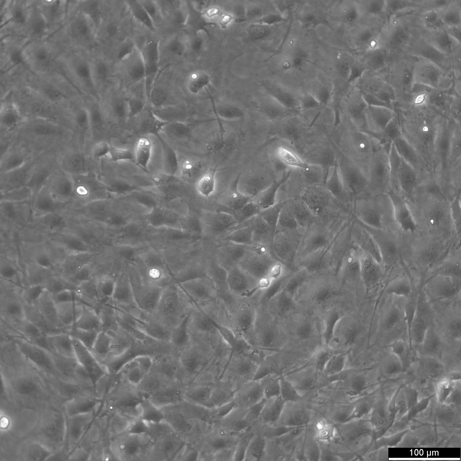
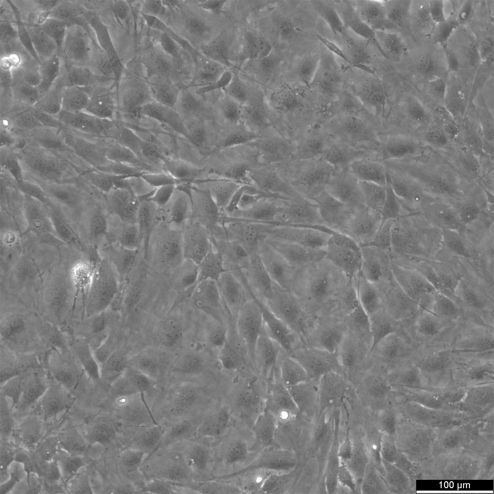
MC3T3-E1 is a clonal, spontaneously immortalized pre-osteoblast cell line of murine origin, assigned Cellosaurus accession CVCL_0409. It was derived from the calvarial bone tissue of a newborn mouse (less than one day old) of unspecified sex. No disease was associated with the donor animal. The cell line was selected on the basis of high ALP activity in the confluent state, a property that presaged its robust capacity for in vitro osteogenesis. In the growing phase, MC3T3-E1 cells display a fibroblastic morphology and form multilayered cultures. Upon induction, they differentiate into osteoblasts, produce a collagenous extracellular matrix, and ultimately give rise to mineralized nodules containing osteocyte-like cells embedded in bone matrix. [1] The doubling time ranges from 24 to 48 hours under standard culture conditions.
SNP array profiling confirmed the C57BL/6 strain origin of MC3T3-E1 cells and has been used to identify karyotypic changes associated with long-term passaging. [3] The line expresses key osteoblast markers including RUNX2, bone sialoprotein, osteocalcin, and osteopontin, particularly in mineralizing subclones. [4] A series of derivative subclones — including MC3T3-E1 Subclone 14 cells and Subclone 4 — were isolated from the parental line and display varying degrees of differentiation and mineralization potential in vitro and in vivo.
Cell Culture Information
- Medium
- Detailed culture conditions are available on the Cytion product page.
- Seeding Density
- See Cytion product page for details.
- Freeze Medium
- See Cytion product page for details.
- Doubling Time
- 24–48 hours
- Growth Type
- Adherent
.jpg)
Advantages of MC3T3-E1 Cells
MC3T3-E1 cells offer a well-defined, reproducible model of the entire osteoblast developmental sequence. Unlike primary calvarial osteoblasts, which are heterogeneous and difficult to maintain, MC3T3-E1 cells are clonally derived and can be expanded consistently across laboratories. Their capacity to progress from proliferating pre-osteoblasts to matrix-depositing osteoblasts and finally to osteocyte-embedded mineralized nodules makes them uniquely suited for temporal studies of osteogenesis. Seminal work by Yohay et al. defined the distinct proliferative and differentiated stages of MC3T3-E1 cells in culture, establishing them as a quantitative in vitro model of osteoblast development. [5] That study demonstrated that MC3T3-E1 cells undergo a predictable sequence of growth arrest, matrix maturation, and mineralization — stages that closely mirror in vivo osteoblast ontogeny.
A further advantage is the availability of well-characterized subclones that provide experimental flexibility. Wang et al. isolated a series of subclones from the parental MC3T3-E1 line, demonstrating that mineralizing subclones selectively express bone sialoprotein, osteocalcin, and PTH/PTHrP receptor mRNAs, while alkaline phosphatase expression appeared in both mineralizing and non-mineralizing subclones. [4] This resource enables researchers to select subclones with defined phenotypes appropriate for their experimental needs. Furthermore, the cells respond robustly to a broad range of osteogenic stimuli — including ascorbic acid, beta-glycerophosphate, BMP-2, dexamethasone, and numerous small molecules — making them ideal for pharmacological and mechanistic studies.
MC3T3-E1 cells are also highly tractable for molecular manipulation. They tolerate transfection, viral transduction, and CRISPR-based gene editing, and they express endogenous signaling pathways (Wnt/β-catenin, BMP/SMAD, PI3K/AKT) that are directly relevant to bone pathophysiology. Their mouse origin facilitates integration with in vivo murine models of osteoporosis and fracture healing, strengthening the translational value of findings obtained in vitro.
Limitations of MC3T3-E1 Cells
Despite their widespread use, MC3T3-E1 cells carry important caveats. As a spontaneously immortalized line, they can accumulate karyotypic abnormalities over extended passaging. SNP array profiling revealed widespread aneuploidy in MC3T3-E1 cultures, and genome-wide changes may affect the reproducibility of differentiation assays at high passage numbers. [3] The original parental line can also undergo spontaneous loss of mineralization capacity, as demonstrated by Baba, who isolated a non-mineralizing subclone (MC3T3-NM4) from the parental MC3T3-E1 population — a finding that underscores the need for careful monitoring of differentiation competence across passages. [6]
The variable osteogenic performance of commercially available MC3T3-E1 subclones represents an additional source of experimental variability. Research has shown that different subclones (e.g., Subclone 4 versus Subclone 14) display markedly different mineralization kinetics and marker gene expression profiles, so direct comparison of results across laboratories using different subclones must be made with caution. Culture medium composition also significantly influences osteogenic outcomes: studies evaluating MC3T3-E1 cells in different culture media reported substantial differences in ALP activity, collagen deposition, and calcification — highlighting the importance of medium standardization in experimental design.
Finally, MC3T3-E1 cells are of murine origin, which limits their direct translational relevance for predicting human osteoblast responses. Species-specific differences in receptor pharmacology, cytokine signaling, and gene regulation mean that findings should be validated in human osteoblast systems before clinical extrapolation. Routine cell line authentication by STR profiling or next-generation sequencing-based methods is strongly recommended to exclude cross-contamination before initiating experiments. [2]
Applications
Osteoblast Differentiation and Osteogenesis Modeling
The most fundamental application of MC3T3-E1 cells is as a quantitative model of osteoblast differentiation. The landmark study by Yohay et al. characterized distinct proliferative and differentiated stages of murine MC3T3-E1 cells in culture, providing a temporal framework for understanding how these pre-osteoblasts transition through growth, matrix maturation, and mineralization phases. [5] That work defined the cell line as a suitable in vitro model of osteogenesis and established experimental benchmarks — such as ALP activity, collagen matrix deposition, and nodule mineralization — that remain in standard use today. The clarity of these developmental stages has made MC3T3-E1 the reference standard against which new osteogenic protocols and differentiation media are evaluated.
Signaling pathway studies have leveraged MC3T3-E1 cells to dissect the molecular machinery of osteogenesis. Wang et al. demonstrated that apelin-13 activates the BMP4/SMAD1/5/8 pathway through its receptor APJ, upregulating osteogenic markers ALP, osteocalcin, osteopontin, and Col1A1, and increasing RUNX2 expression at both mRNA and protein levels in MC3T3-E1 cells. [7] MicroRNA regulation of differentiation has also been explored: Zhai et al. showed that the miR-211-5p/FOXO3 axis promotes osteogenic differentiation of MC3T3-E1 cells and BMSCs by activating the Wnt/β-catenin pathway, with implications for osteoporotic fracture healing. [8] Fluid shear stress research using MC3T3-E1 cells further revealed that AnnexinA6-mediated autophagy regulates mechanically induced osteogenic differentiation, a finding relevant to understanding load-bearing bone physiology. [9]
MC3T3-E1 cells have been used to form three-dimensional spheroids that better recapitulate the in vivo osteoblast microenvironment. Wen et al. generated spontaneous MC3T3-E1 spheroids using a low-adhesion culture system and demonstrated significantly enhanced ALP activity and osteogenic gene expression compared to monolayer cultures. [10] These spheroids promoted alveolar bone regeneration and suppressed inflammation in a murine tooth autotransplantation model, highlighting the value of three-dimensional MC3T3-E1 culture systems for tissue engineering applications.
Osteoporosis Research and Drug Discovery
MC3T3-E1 cells serve as a primary in vitro platform for evaluating candidate therapeutics against osteoporosis. Quercetin, a naturally occurring flavonoid, was shown to mitigate osteoblast dysfunction in osteoporotic rats by regulating ferroptosis through the Nrf2/SLC7A11/GPX4 signaling pathway; in vitro validation using MC3T3-E1 cells confirmed the pathway's role in osteoblast survival and function. [11] Similarly, pilose antler protein extract (PAE) promoted MC3T3-E1 cell viability, proliferation, and osteogenic differentiation while activating the Wnt/β-catenin signaling pathway, mirroring its beneficial effects on trabecular microarchitecture in ovariectomized rats. [12]
Polysaccharides from traditional medicinal plants have also been evaluated using MC3T3-E1 cells as the in vitro component of a broader anti-osteoporosis investigation. Yang et al. characterized two Psoralea corylifolia polysaccharides and demonstrated their capacity to stimulate osteogenic activity in MC3T3-E1 cells, complementing in vivo findings of improved bone mineral density in glucocorticoid-induced osteoporotic mice. [13] Curcumin nanoparticles combined with narlumosbart were shown to synergistically enhance osteoblast differentiation in MC3T3-E1 cells via the Wnt/β-catenin pathway, offering a combinatorial nanoparticle strategy for bone trauma repair. [14]
Osteomyelitis, a severe bone infection, has been studied using MC3T3-E1 cells treated with staphylococcal protein A (SPA) as an in vitro infection model. Chen et al. used Mendelian randomization combined with these in vitro experiments to demonstrate that the circulating metabolite p-cresol sulfate impairs MC3T3-E1 cell proliferation and osteogenic differentiation, providing new mechanistic insights into metabolic drivers of osteomyelitis pathogenesis. [15] Ferroptosis inhibition using ferrostatin-1 has also been investigated in MC3T3-E1 cells, with results suggesting that suppressing ferroptotic cell death enhances osteoblast viability and bone-forming activity.
Biomaterials, Implant Science, and Bone Tissue Engineering
MC3T3-E1 cells are the reference standard for assessing the biocompatibility and osteoinductive potential of novel biomaterials. Liu et al. developed a terbium-doped 3D-printed carbonate hydroxyapatite (Tb-CHA) scaffold and used MC3T3-E1 cells to demonstrate that the material significantly enhanced cell proliferation and differentiation in vitro, with subsequent in vivo validation in a rat calvarial defect model confirming its osteogenic and angiogenic capabilities. [16] The ability to correlate MC3T3-E1 in vitro responses with in vivo bone regeneration outcomes makes this cell line especially valuable in the iterative design of bone substitute materials.
Titanium implant surface engineering has relied heavily on MC3T3-E1 cells to quantify osseointegration potential. Liao and Li fabricated a composite coating of epigallocatechin-3-gallate (EGCG), hydroxyapatite (HAP), and silver nanoparticles (AgNPs) on titanium surfaces and showed that this EGCG/HAP/AgNPs coating simultaneously improved antibacterial activity and promoted MC3T3-E1 cell adhesion, proliferation, and osteogenic differentiation. [17] These findings illustrate how MC3T3-E1 cells help optimize implant coatings that must balance antimicrobial efficacy with host cell compatibility.
Zinc oxide nanoparticles (ZnO-NPs) represent another class of biomedical materials evaluated in MC3T3-E1 cells. Ryu et al. systematically assessed the concentration-dependent effects of ZnO-NP extracts on cytocompatibility and osteogenic activity, finding that low concentrations promoted osteoblastic responses while higher concentrations were cytotoxic. [18] This dose-dependent characterization underscores the importance of using standardized MC3T3-E1 assays when establishing the safety and efficacy windows of nanomaterial-based bone repair strategies. Cold atmospheric plasma (CAP) technology has similarly been assessed for its capacity to stimulate MC3T3-E1 osteogenic differentiation in a time-dependent manner, opening potential applications in alveolar bone regeneration. [19]
Microgravity and Mechanobiology
MC3T3-E1 cells have contributed foundational insights into how mechanical and gravitational unloading affects osteoblast biology. Hughes-Fulford and Lewis launched MC3T3-E1 osteoblasts aboard the STS-56 Space Shuttle mission and demonstrated that cells activated in microgravity showed impaired growth despite normal serum stimulation, with altered cytoskeletal organization and reduced prostaglandin synthesis compared with ground controls. [20] These findings provided a cellular mechanistic basis for the well-documented loss of bone mineral density experienced by astronauts during long-duration spaceflight.
Subsequent work by Hughes-Fulford extended these observations to gene expression and signal transduction, demonstrating that microgravity alters the transcriptional program of MC3T3-E1 osteoblasts in ways that help explain reduced bone formation in the absence of gravitational load. [21] Together, these space biology studies established MC3T3-E1 cells as the principal in vitro model for investigating the cellular consequences of microgravity on the skeleton, with direct relevance to developing countermeasures for bone loss in space medicine.
On Earth, mechanobiology studies have exploited the MC3T3-E1 system to understand how fluid shear stress — a proxy for interstitial flow in bone lacunocanalicular networks — regulates osteoblast differentiation. Research demonstrated that autophagy mediated by AnnexinA6 links mechanical stimulation to osteogenic gene expression in MC3T3-E1 cells, providing molecular detail on how bone cells sense and respond to physical loading. [9] These mechanobiology applications position MC3T3-E1 cells at the interface of biophysics and regenerative medicine.
Periodontal and Alveolar Bone Regeneration
Periodontitis-related bone loss is a major clinical challenge, and MC3T3-E1 cells have become a key in vitro tool for testing periodontal regenerative strategies. Kong et al. developed a multifunctional carbon dot nanomaterial (ASA-ALN-CDs) derived from aspirin and alendronate sodium that demonstrated potent antibiofilm activity against periodontal pathogens and promoted MC3T3-E1 osteogenic differentiation under inflammatory conditions. [22] In a rat periodontitis model, the material reduced alveolar bone loss and inflammation by inhibiting the HIF-1α signaling pathway, with the MC3T3-E1 in vitro data providing direct mechanistic support for the observed in vivo effects.
Cold atmospheric plasma studies specifically targeted alveolar bone regeneration by exposing MC3T3-E1 cells to a custom helium CAP source. Negrescu et al. showed that CAP treatment modulated cell survival and osteogenic differentiation in a time-dependent manner, assessed through live/dead assays, CCK-8 viability assays, ALP activity measurements, and morphological characterization. [19] These results positioned CAP as a candidate physical adjunct therapy for alveolar bone defects, with MC3T3-E1 cells providing the quantitative differentiation readouts needed to optimize treatment parameters.
The three-dimensional spheroid culture approach developed with MC3T3-E1 cells has direct relevance to dental regenerative medicine. The spheroid-collagen scaffold system evaluated by Wen et al. in a tooth autotransplantation model achieved superior alveolar bone regeneration and anti-inflammatory gene expression compared to monolayer cell controls, suggesting that the enhanced paracrine activity of MC3T3-E1 spheroids may translate into clinically meaningful gains in dental socket healing. [10]
Conclusion
MC3T3-E1 cells remain an indispensable and extensively validated model for bone biology research. From their original characterization as a clonal pre-osteoblast line capable of in vitro mineralization, to their current deployment in cutting-edge fields including 3D bioprinting, nanoparticle-based drug delivery, periodontal regeneration, and space medicine, these cells have demonstrated extraordinary scientific versatility. Their well-defined differentiation stages, robust marker expression, and responsiveness to a wide range of osteogenic stimuli make them the benchmark cell line for evaluating new biomaterials, identifying therapeutic targets in osteoporosis and osteomyelitis, and dissecting the signaling pathways that govern bone formation. Whether you are investigating ferroptosis, mechanobiology, or next-generation implant coatings, MC3T3-E1 cells provide the reproducible, biologically relevant platform your research demands. Explore the full specifications or order MC3T3-E1 Cells directly at cytion.com.
Key Publications
- Sudo H, Kodama HA, Amagai Y (1983) In vitro differentiation and calcification in a new clonal osteogenic cell line derived from newborn mouse calvaria. The Journal of Cell Biology. PMID: 6826647
- Chen YH, Connelly JP, Florian C (2023) Short tandem repeat profiling via next-generation sequencing for cell line authentication. Disease Models & Mechanisms. PMID: 37712227
- Didion JP, Buus RJ, Naghashfar Z (2014) SNP array profiling of mouse cell lines identifies their strains of origin and reveals cross-contamination and widespread aneuploidy. BMC Genomics. PMID: 25277546
- Wang D, Christensen K, Chawla K (1999) Isolation and characterization of MC3T3-E1 preosteoblast subclones with distinct in vitro and in vivo differentiation/mineralization potential. Journal of Bone and Mineral Research. PMID: 10352097
- Quarles LD, Yohay DA, Lever LW (1992) Distinct proliferative and differentiated stages of murine MC3T3-E1 cells in culture: An in vitro model of osteoblast development. Journal of Bone and Mineral Research. PMID: 1414487
- Baba TT (2000) Restoration of mineral depositions by dexamethasone in the matrix of nonmineralizing osteoblastic cells subcloned from MC3T3-E1 cells. Calcified Tissue International. PMID: 11136541
- Wang C, Li Z, Yang H (2026) Apelin-13 activates the BMP4/SMAD pathway through APJ to enhance osteoblastic differentiation and mineralization. Tissue & Cell. PMID: 42269418
- Zhai H, Lin M, Lu C (2026) miR-211-5p/FOXO3 axis accelerates osteogenic differentiation and fracture healing by mediating Wnt/β-catenin pathway. Journal of Orthopaedic Surgery and Research. PMID: 42219493
- Pei T, Su G, Yang J (2026) Correction: Fluid Shear Stress Regulates Osteogenic Differentiation via AnnexinA6-Mediated Autophagy in MC3T3-E1 Cells. International Journal of Molecular Sciences. PMID: 42196630
- Wen Z, Li X, Yue L (2026) Spontaneously formed mesenchymal stem cell spheroids enhance alveolar bone regeneration and suppress inflammation following tooth autotransplantation. Regenerative Therapy. PMID: 42199333
- Gong J, Ma Y, Huang J (2026) Pilose Antler Protein Extract Alleviates Osteoporosis and Is Associated with Activation of the Wnt/β-Catenin Signaling Pathway. Pharmaceuticals (Basel, Switzerland). PMID: 42198339
- He L, Zheng Y, Zeng Z (2026) Effect of quercetin on osteoblast dysfunction in osteoporotic rats by regulating Nrf2/SLC7A11/GPX4 pathway-mediated ferroptosis. Cytotechnology. PMID: 42238059
- Yang J, Tang Z, Chen C (2026) Structural characterization and in vivo and in vitro anti-osteoporosis potential of two Psoralea corylifolia L. polysaccharides. Carbohydrate Polymers. PMID: 42173575
- Zhang H, Zhao Y (2026) Curcumin nanoparticles combined with narlumosbart regulate wingless-type MMTV integration site (wnt)/β-catenin signaling pathway in osteoblast-like cells. Pakistan Journal of Pharmaceutical Sciences. PMID: 42170973
- Chen X, Mo R, Yang S (2026) Inflammatory cytokines and metabolic pathways in osteomyelitis: Mendelian randomization insights and experimental validation. Advances in Clinical and Experimental Medicine. PMID: 42200571
- Liu P, Shen J, Bian Y (2026) Terbium-doped 3D-printed carbonate hydroxyapatite scaffold enhances bone regeneration. Journal of Translational Medicine. PMID: 42260646
- Liao J, Li Z (2026) Antibacterial and osteogenic effect of EGCG/HAP/AgNPs composite coating on titanium surface. Journal of Materials Science: Materials in Medicine. PMID: 42216990
- Ryu JH, Mangal U, Kim JH (2026) Dose-dependent effects of zinc oxide nanoparticles on pre-osteoblast cellular response. Scientific Reports. PMID: 42303653
- Negrescu AM, Zampieri L, Martines E (2026) Time-dependent modulation of MC3T3-E1 pre-osteoblasts by a helium cold plasma: a preliminary study on its potential relevance for alveolar bone regeneration. Biomedical Physics & Engineering Express. PMID: 42208566
- Kong J, Li H, Guo X (2026) Therapeutic Efficacy of ASA-ALN-CDs in Periodontitis: From Antibiofilm/Anti-Inflammation to Alveolar Bone Regeneration. ACS Biomaterials Science & Engineering. PMID: 42213513
- Hughes-Fulford M, Lewis ML (1996) Effects of microgravity on osteoblast growth activation. Experimental Cell Research. PMID: 8612673
- Hughes-Fulford M (2001) Changes in gene expression and signal transduction in microgravity. Journal of Gravitational Physiology. PMID: 12638602
Explore the full specifications or order MC3T3-E1 Cells at cytion.com.