
Published: 17 October 2023 | Last reviewed: May 2026
A431 Cells: Origin, Culture Conditions, and Research Applications
A431 is a human epidermoid carcinoma cell line derived from the skin of an 85-year-old female patient. It is one of the most widely used cell lines in biomedical research, particularly in cancer biology, immuno-oncology, dermatology, and drug discovery. A431 cells massively overexpress the epidermal growth factor receptor (EGFR) — carrying approximately 2–3 million receptors per cell, roughly 20–50 times more than normal epithelial cells — making them the gold-standard in vitro model for EGFR signalling studies and the testing of EGFR-targeted therapies such as cetuximab and erlotinib. A431 cells are available exclusively from Cytion, your independent European cell bank.
- Species / Origin
Human (female, 85 years old) - Tissue
Skin epidermis (epidermoid carcinoma) - Morphology
Epithelial, adherent - Doubling Time
80–100 hours - Growth Medium
DMEM + 10% FBS - Biosafety Level
BSL-1 - Key Feature
EGFR overexpression (~2–3M receptors/cell) - Key Biomarkers
EGFR (amplified), TP53 (mutant), KRAS (wild-type), PTEN (expressed) - Product Number
300112 — Order A431 Cells from Cytion
- A431 cells: General characteristics and origin
- A431 cell line: Culturing information
- Advantages & disadvantages of A431 cell line
- Research applications of A431 cells
- A431 vs. other skin cancer cell lines
- Research publications featuring A431 cells
- Resources, protocols, and videos
- Frequently asked questions
A431 cells: General characteristics and origin
This section covers the key biological properties of the A431 cell line — including its origin, morphology, karyotype, and the EGFR overexpression that makes it uniquely valuable for research.
A431 Cell Line Origin and History
- A431, a human skin cancer cell line, was obtained from the epidermis of an 85-year-old female epidermoid carcinoma patient [1]. It was established by D.J. Giard et al., who developed several other cell lines from solid tumours.
- The cell line has been widely distributed and validated by independent cell banks worldwide, including Cytion, as a reliable and reproducible research model.
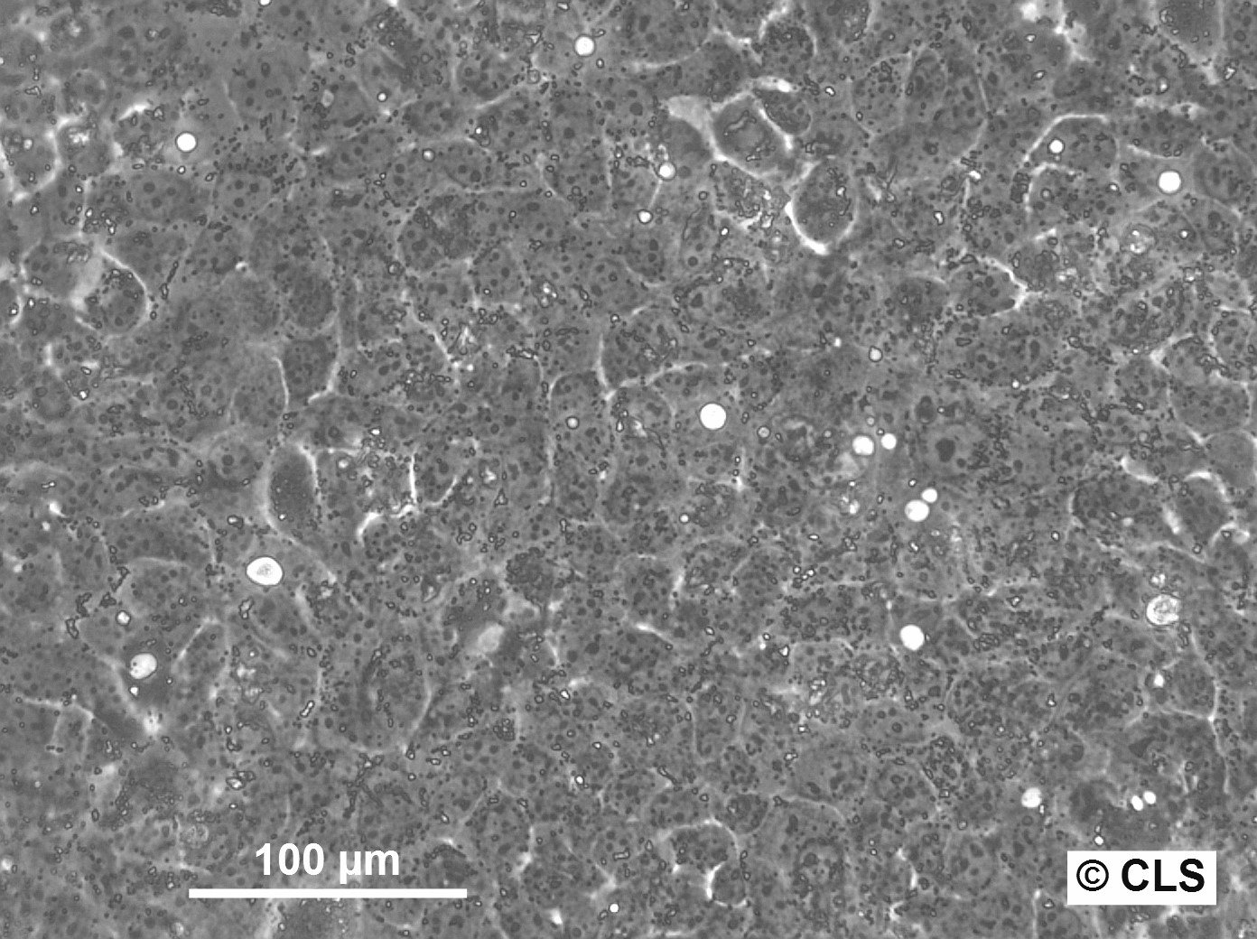
A431 Morphology and Karyotype
- A-431 cells possess epithelial morphology. They aggregate and form cell clusters under standard culture conditions.
- The A431 skin cancer cell line is hypertriploid. The modal chromosome number is 74, occurring in approximately 36% of cells. Higher ploidies are also present in around 1% of the cell population.
EGFR Expression in A431 Cells
EGFR overexpression is the defining characteristic of A431 cells and the primary reason for their widespread use. A431 cells carry a genomic amplification of the EGFR gene locus, resulting in approximately 2–3 million EGFR receptors per cell — 20 to 50 times higher than normal epithelial cells. This makes A431 the gold-standard positive control for:
- EGFR antibody validation by Western blot, flow cytometry, and immunofluorescence
- Testing EGFR-targeted monoclonal antibodies such as cetuximab and panitumumab
- Evaluating EGFR tyrosine kinase inhibitors (TKIs) such as erlotinib, gefitinib, and afatinib
- Studying downstream signalling pathways including PI3K/AKT/mTOR and RAS/MAPK/ERK
A431 cell line: Culturing information
Knowing a cell line's culture requirements makes handling trouble-free. This section covers the doubling time, growth medium, passaging, cryopreservation, and biosafety requirements for the A431 cell line.
Key Points for Culturing A431 Cells
Doubling Time:
The population doubling time for A431 cells ranges from 80 to 100 hours.
Adherent or in Suspension:
A431 is an adherent cell line.
Seeding Density:
1 x 10⁴ cells/cm2 is ideal for the A431 cell line. Cells reach confluence in approximately 4 days at this density. Adherent cells are washed with PBS (1×) and incubated with Accutase passaging solution, then resuspended in culture medium, centrifuged, and dispensed into new flasks.
Growth Medium:
DMEM medium supplemented with 10% fetal bovine serum (FBS), 4.5 g/L Glucose, 1.0 mM Sodium pyruvate, 1.5 g/L NaHCO3, and 4 mM L-Glutamine. Media should be replaced every 2–3 days.
Growth Conditions:
A431 cancer cells are grown in a humidified incubator with 5% CO2 at 37°C.
Storage:
Store in an electric freezer or vapour phase of liquid nitrogen at below −150°C to protect cell viability.
Freezing Process and Medium:
CM-1 or CM-ACF are the recommended freezing media for A431 cells. Use a controlled-rate slow freezing method (−1°C/min).
Thawing Process:
Thaw frozen A431 cells rapidly in a 37°C water bath (40–60 seconds). When a small ice clump remains, add culture medium and centrifuge. Resuspend harvested cells and transfer to culture flasks.
Biosafety Level:
Biosafety Level 1 (BSL-1) is recommended for handling A431 cultures.

Advantages & disadvantages of A431 cell line
The A431 cell line possesses distinguishable characteristics that make it both highly useful and contextually limited. Here is a balanced summary.
Advantages
The main advantages of the A431 cancer cell line are:
EGFR overexpression
A431 cells overexpress EGFR at 2–3 million receptors per cell, serving as the gold-standard positive control for EGFR signalling studies and anti-EGFR drug validation (cetuximab, erlotinib, gefitinib).
Tumorigenicity
A431 cells are tumorigenic and readily form tumours in immunocompromised mice, making them a reliable tool for xenograft cancer models to study tumour growth dynamics and assess novel cancer treatments in vivo.
Well characterised
Decades of published research mean that A431 cells are extensively characterised at the genomic, proteomic, and phenotypic level, providing a robust scientific foundation for experimental design.
3D spheroid formation
A431 cells readily form compact 3D spheroids under ultra-low attachment conditions, making them compatible with modern organoid and spheroid screening platforms for drug penetration and efficacy studies.
Disadvantages
The disadvantages of A431 cells are:
Genetic abnormalities
A431 is a cancer cell line with accumulated genetic mutations and chromosomal alterations (hypertriploid, modal number 74) that may not fully recapitulate the original tumour characteristics.
Microbial contamination risk
A431 is prone to bacterial contamination. Maintaining strict aseptic technique and routine mycoplasma testing are essential for reliable results.
Not representative of all EGFR tumours
Because EGFR expression in A431 is driven by gene amplification rather than mutation (as in lung cancer), findings may not directly translate to EGFR-mutant tumour contexts such as non-small-cell lung cancer (NSCLC).
Research applications of A431 cells
The A431 cell line is extensively used across multiple research disciplines. Its unique EGFR biology, tumorigenicity, and well-characterised genome make it one of the most versatile human cancer cell lines available.
- Cancer biology: A431 is a powerful tool for investigating cellular and molecular mechanisms driving cancer growth, metastasis, and invasion. Studies have mapped PI3K/AKT/mTOR and RAS/MAPK/ERK signalling in this cell line, with inhibition shown to induce apoptosis in A431 skin tumour cells [2, 3].
- EGFR-targeted therapy testing: A431 cells are the reference standard for evaluating anti-EGFR therapeutics — monoclonal antibodies (cetuximab, panitumumab) and small-molecule TKIs (erlotinib, gefitinib, afatinib, osimertinib).
- Drug testing and evaluation: A431 cells are used to evaluate novel anti-cancer drug candidates. A 2022 study by Rahim et al. demonstrated potent antiproliferative effects of biogenically synthesised silver nanoparticles from Alstonia angustiloba on A431 cells [4].
- Tumour xenograft models: A431 cells are tumorigenic in immunodeficient mice, enabling subcutaneous and orthotopic xenograft models for in vivo efficacy studies. Lim et al. used A431-derived xenografts to evaluate the radiosensitising effects of exogenous EGF in vivo [5].
- Skin toxicity and dermatology research: A431 cells are widely used by pharmaceutical and cosmetic companies to assess dermal toxicity, skin penetration, and safety profiles of topically applied compounds.
- 3D spheroid and organoid models: A431 cells form compact 3D spheroids under ultra-low attachment or hanging-drop conditions, enabling more physiologically relevant drug penetration and resistance studies.
- EGFR antibody validation: Due to their extreme EGFR density, A431 cells are the standard positive control for validating anti-EGFR antibodies in Western blot, immunofluorescence, IHC, and flow cytometry.
A431 vs. other skin cancer cell lines
Choosing the right cell line is critical for experimental validity. Here is how A431 compares to other commonly used skin and squamous cancer cell lines:
| Feature | A431 | A375 (Melanoma) | SCC-25 (Oral SCC) | HaCaT (Keratinocyte) |
|---|---|---|---|---|
| Cancer type | Epidermoid carcinoma | Malignant melanoma | Oral squamous cell carcinoma | Immortalised keratinocyte |
| EGFR expression | +++ (~2–3M/cell) | + (low) | ++ (moderate) | ++ (moderate) |
| Tumorigenicity | Yes | Yes | Yes | No |
| Morphology | Epithelial, adherent | Spindle-shaped, adherent | Epithelial, adherent | Epithelial, adherent |
| BSL | 1 | 1 | 1 | 1 |
| Primary use | EGFR research, anti-cancer drug testing | Melanoma biology, BRAF inhibitors | Head & neck cancer, radiation studies | Skin biology, barrier function |
Research Publications Featuring A431 Cells
Here are some significant peer-reviewed publications featuring the A431 skin carcinoma cell line.
Published in Materials Today Communications (2020). Evaluated the anticancer potential of poly-ε-caprolactone curcumin-loaded nanofibers and polyvinyl alcohol-AuNPs against A431 cancer cells.
miRNA-221 promotes cutaneous squamous cell carcinoma progression by targeting PTEN
Published in Cellular & Molecular Biology Letters (2019). Proposed that microRNA-221 plays an oncogenic role in cutaneous squamous cell carcinoma by targeting the PTEN tumour suppressor gene.
Published in BMC Complementary Medicine and Therapies (2016). Showed that vincristine from the fungus Eutypella spp–CrP14 induces apoptosis in A431 cells.
Overexpression of CDC42SE1 in A431 cells reduced cell proliferation by inhibiting the Akt pathway
Published in Cells (2019). Identified CDC42SE1 as a potential biomarker of skin cancer progression, with its downregulation facilitating tumorigenesis in A431 cells.
Metformin inhibits the proliferation of A431 cells by modulating the PI3K/Akt signaling pathway
Published in Experimental and Therapeutic Medicine (2015). Demonstrated that metformin suppresses A431 proliferation by regulating the PI3K/Akt signalling pathway.
Resources for A431 Cell line: Protocols, Videos, and More
The following resources provide practical guidance for working with A431 cells in the laboratory.
- Transfecting A-431 cells — A step-by-step video guide to transfecting A431 cancer cells.
- A431 cancer cell line — Cytion product page — Full cell culture information: growth medium, seeding density, passaging, cryopreservation, and quality certificates.
- A431 cell culturing protocol — A concise external protocol for culturing the A431 cell line from Bio-Protocol.
- Browse all skin cancer cell lines from Cytion — Explore the full range of skin cancer cell lines available from Cytion.